Evidence collection21 October 2020
Published 20 April 2020 | Updated 31 October 2025
Introduction
COVID-19 is the official designation of the human disease caused by the novel Coronavirus (SARS-CoV-2, Severe Acute Respiratory Syndrome Corona Virus2) first identified in Wuhan China in December 2019.
Coronaviruses are a large family of RNA viruses that are commonly found in humans as well as other mammals, birds, and reptiles. They all have a characteristic crown (‘corona’) of protein spikes around their lipid envelope. However, the common coronaviruses causing respiratory or gastrointestinal disease in our veterinary patients are alpha coronaviruses, whereas the SARS-CoV-2, is a beta coronavirus, closely related to the viruses that cause SARS and MERS.
Genomic analyses of SARS-CoV-2 indicated that mutations of the receptor binding domain (RBD) of the spike protein present on the surface of the virus optimised the ability to bind to angiotensin-converting enzyme 2 (ACE2) receptors present on the surface of human cells. It has been suggested that SARS-CoV-2 originated either through natural selection in an animal host before zoonotic transfer or as a result of natural selection in humans following zoonotic transfer1.
A recently published paper identified key interactions between spike protein and host receptor ACE2, as the means of both the cross-species and human-to-human transmissions of SARS-CoV (the virus that causes SARS). The authors predict that SARS-CoV-2 also uses ACE2 as its receptor and that the virus could also bind to ACE2 in pigs, ferret, cats and some non-human primates with similar efficiency as it does in people2.
There are three ways that animals could be involved in COVID-19:
- They could be contaminated with live virus and act as fomites
- They could become infected with SARS-CoV-2 and develop signs of infection
- They could become infected and pass on the virus to other animals or humans.
Current evidence indicates that the predominant route of transmission of COVID-19 is from human to human. However, there are now reports of a small number of companion animals becoming contaminated or infected after close contact with infected people. The World Organisation for Animal Health (OIE) reports that studies are underway to better understand the susceptibility of different animal species to the COVID-19 virus and to assess infection dynamics in susceptible animal species, but that there is no evidence to suggest that animals infected by humans are playing a role in the spread of COVID-19. Human outbreaks are driven by person to person contact3.
It should be noted that when reviewing case reports and published papers three different types of tests are referred to:
PCR (Polymerase Chain Reaction): this test detects the presence of viral genetic material (in this case RNA) but cannot distinguish between infection and contamination.
Antibody tests, which can include Enzyme-Linked Immunosorbent Assays (ELISAs) and Virus Neutralization Tests: detect the presence of antibody to the virus. A positive antibody test indicates that the animal has mounted an immune response to the virus, but does not give any information on whether the animal was clinically ill or has shed the virus at any time.
Virus isolation: used to test for the presence of live virus, and a positive test means that an animal has the potential to shed live virus.
References
- Anderson, Kristian G et al (2020) The proximal origin of SARS-CoV-2 Nature Medicine 26 pp 450-452 https://doi.org/10.1038/s41591-020-0820-9
- Wan, Yushan et al. (2020) Receptor recognition by novel coronavirus from Wuhan: an analysis based on decade-long structural studies of SARS. Journal of Virology 2020 94 (7) e00127-20 https://doi.org/10.1128/JVI.00127-20
- OIE (2020) Questions and Answers on the 2019 Coronavirus Disease (COVID-19) [online] Available at https://www.woah.org/fileadmin/Home/MM/A_COVID-27.11.2020.pdf. [Accessed 23 September 2025]
Case reports
There have been a small number of reports of animals being infected with SARS-CoV-2. To date, all these animals appear to have been infected through contact with infected people. There is no evidence of humans being infected through contact with domestic animals.
There continue to be further case reports of pet animals infected with SARS-CoV-2, in all cases these appear to come from households in which at least one person is infected with COVID-19.
A full list of case reports of confirmed infection of SARS-CoV-2 in animals (Events in animals tab) can be found on the OIE website.
SARS-ANI VIS: a global open access dataset of reported SARS-CoV-2 events in animals. [Complexity Science Hub, Vienna] [online]. Available from: https://vis.csh.ac.at/sars-ani/#infections [Accessed 10 August 2022].
Variants (Delta and Alpha)
- Tewari, D. et al. (2023) SARS-CoV-2 infection dynamics in the Pittsburgh Zoo wild felids with two viral variants (Delta and Alpha) during the 2021–2022 pandemic in the United States. Animals, 13 (19), no. 3094. https://doi.org/10.3390/ani13193094
Variant (Omicron)
- Klein, C. et al. (2023) Dogs and cats are less susceptible to the Omicron variant of concern of SARS-CoV-2: A field study in Germany, 2021/2022. Transboundary and Emerging Diseases, 2023, no. 1868732. https://doi.org/10.1155/2023/1868732
Variant B.1.1.529 (Omicron)
- Sánchez-Morales, L. et al. (2022) The Omicron (B.1.1.529) SARS-CoV-2 variant of concern also affects companion animals. Frontiers in Veterinary Science, 9, no. 940710. https://www.frontiersin.org/articles/10.3389/fvets.2022.940710
Variant B.1.617.2 (Delta)
- Barroso-Arevalo, S. et al (2022) First detection of SARS-CoV-2 B.1.617.2 (Delta) Variant of Concern in a symptomatic cat in Spain. Frontiers in Veterinary Science, 9, no. 841430. https://doi.org/10.3389/fvets.2022.841430
- Wendling, N.M. et al (2022) Transmission of SARS‐CoV‐2 Delta variant (B.1.617.2) from a fully vaccinated human to a canine in Georgia, July 2021. Zoonoses and Public Health. https://doi.org/10.1111/zph.12944
- Lenz, O.C. et al (2022) SARS-CoV-2 Delta Variant (AY.3) in the feces of a domestic cat. Viruses, 14 (2), p. 421. https://doi.org/10.3390/v14020421
Variant B.1.1.7 (Alpha)
Case reports reporting infection of cats and dogs with alpha variant (B.1.1.7.)
- Pecora, A. et al (2022) Anthropogenic infection of domestic cats with SARS-CoV-2 Alpha Variant B.1.1.7 lineage in Buenos Aires. Frontiers in Veterinary Science, 9, no. 790058. https://doi.org/10.3389/fvets.2022.790058
- Keller, M. et al (2021) Detection of SARS-CoV-2 variant B. 1.1. 7 in a cat in Germany. Research in Veterinary Science, 140, pp. 229-232. https://doi.org/10.1016/j.rvsc.2021.09.008
- Curukoglu, A. et al (2021) First direct human‐to‐cat transmission of the SARS‐CoV‐2 B. 1.1. 7 variant. Australian Veterinary Journal, 99 (11), pp. 482-488. https://doi.org/10.1111/avj.13109
- Miró, G. et al (2021) SARS-CoV-2 infection in one cat and three dogs living in COVID-19-positive households in Madrid, Spain. Frontiers in Veterinary Science, p. 1292. https://doi.org/10.3389/fvets.2021.779341
- Chetboul, V. et al ( 2021) Myocarditis and subclinical-like infection associated with SARS-CoV-2 in two cats living in the same household in France: a case report with literature review. Frontiers in Veterinary Science, 8, p. 1174. https://doi.org/10.3389/fvets.2021.748869
- Hamer, S.A. et al (2021) SARS‐CoV‐2 B.1.1.7 variant of concern detected in a pet dog and cat after exposure to a person with COVID‐19, USA. Transboundary and Emerging Diseases. https://doi.org/10.1111/tbed.14122
Hamsters Hong Kong
- Government of the Hong Kong Special Administrative Region: Agriculture, Fisheries and Conservation Department (2022) Press release: hamster samples preliminarily test positive for COVID-19 virus [online] . Available at: https://www.afcd.gov.hk/english/publications/publications_press/pr2516.html [Accessed 29 January 2022]
There have been multiple reports in the news of hamsters from a pet shop in Hong Kong being culled, and that other pet shops selling hamsters must suspend business, over fears of the spread of COVID.
While it has been known for some time that hamsters can be experimentally infected with SARS-CoV-2 and spread the virus to other hamsters, to date there have not been any reports of natural infection or any evidence that they can transmit the virus to other species.
The Agriculture, Fisheries and Conservation Department (AFCD) in Hong Kong have released the following statement, which reports that 11 hamsters have so far given preliminary positive tests for SARS-CoV-2, while all other animals tested have tested negative.
From pet hamsters to humans
- Yen, H.L. (2022) Transmission of SARS-CoV-2 (variant Delta) from pet hamsters to humans and onward human propagation of the adapted strain: a case study. Preprints with The Lancet https://dx.doi.org/10.2139/ssrn.4017393
This paper, currently in preprint, reports on the investigation of SARS-CoV-2 infection in hamsters at a pet shop in Hong Kong and provides some preliminary evidence of transmission of infection from hamsters to humans.
The first human case was a 23-year-old female vaccinated pet shop worker (Patient 1), presented with sore throat and cough, confirmed to be infection with COVID (VOC Delta -AY127 virus lineage). She has no known contact with infected humans.
A mother (patient 2) and her daughter who visited the pet shop and purchased a hamster, also tested positive for SARS-CoV-2 on PCR tests. Subsequently two other members of the household were also confirmed to be infected.
Initial screening of the animals in the pet shop, hamsters (n=69), rabbits (n=42) and Guinea pigs (n=14) returned seven (10.2%) positive swabs from hamsters while none of those from other animals tested positive by RT-PCR. The wholesale warehouse supplying this pet-shop chain was then investigated, with 511 swabs collected from hamsters (n=137), rabbits (n=204), Guinea pigs (n=52), chinchilla (n=116) and mice (n=2) housed there. Here one Syrian hamster swab was RT-PCR positive for SARS-CoV-2.
Since the initial screening suggested that hamsters were infected at both the warehouse and the pet shop, a more detailed sampling was carried out at both sites with swabs and serum being collected from the Syrian and dwarf hamsters.
At the pet shop 8 (50%) of 16 Syrian hamsters had evidence of infection, either by serology or confirmed RT-PCR, with 4 animals testing positive by both serology and RT-PCR, 3 animals tested positive by RT-PCR alone and 1 animal tested positive by serology alone. A total of 3 cages housing Syrian hamsters were sampled and two (66.7%) had animals with confirmed RT-PCR or serology results. In contrast, none of 20 cages housing dwarf hamsters were positive in either RT-PCR or antibody assays.
At the warehouse twelve Syrian hamsters and 55 dwarf hamsters were sampled. Two (16.7%) of the swabs were RT-PCR positive and seven (58.3%) of the sera from Syrian hamsters, had evidence of antibody. The authors interpreted the detection of 5 animals with antibodies but without viral RNA to suggest that infection may have occurred at an earlier date.
Investigation into the source of the infected hamsters suggested that they were imported from Netherlands to Hong Kong in two different batches (arrival dates: 22-December-2021 and 7-January-2022) and that some hamsters arriving on the 7-January-2022 were transferred to pet shop A on the day of arrival.
The virus sequenced from the hamsters were genetically closely related to recent AY.127 viruses detected in multiple European countries. By contrast, none of the AY.127 sequences previously detected from returning travellers in Hong Kong is genetically similar to the sequences detected in this outbreak. This further supports the hypothesis that this outbreak was caused by a recent introduction of AY.127 virus from Europe.
Specimens from the first 3 human cases (Patients 1-3) and positive hamster samples collected in pet shop A (n=11) and the warehouse (n=1) were subjected to full viral genome sequence analysis. The viral genomes all belong to the Delta AY.127 viral lineage.
While the sequences from these human and hamster cases were highly similar, they were not identical. The divergent date of this cluster of human and hamster viruses is estimated to be on 21-November-2021 (; 95% CI range: 18-October-2021 to 16- December-2021). Interestingly, the viral genome of the pet shop worker (patient 1) was phylogenetically distinct (5 nucleotides different) from those of the mother (patient 2) and her husband (patient 3), which were identical.
The authors considered that these results suggest that Patient 1 and Patient 2 acquired the infection independently from hamsters at the pet shop rather than from each other. As Patient 3 did not visit the pet shop, these findings further suggest that the SARS-CoV-2 virus circulating in hamsters allowed at least 1 human-to-human transmission.
The authors conclude that pet hamsters can acquire SARS-CoV-2 infection in real-life settings and can transmit the virus back to humans.
Please note this paper has been published as a preprint and has not been subject to peer review.
Further information
For anyone looking for further details of the pathogenesis and signs of SARS-CoV-2 in hamsters, the following two papers may be of interest.
- Sia, S.F. et al (2020) Pathogenesis and transmission of SARS-CoV-2 in golden hamsters. Nature, 583 (7818), pp. 834-838. https://doi.org/10.1038/s41586-020-2342-5
This paper reports on the presence of viral antigens in nasal mucosa, bronchial epithelial cells and areas of lung consolidation on days 2 and 5 after inoculation with SARS-CoV-2, followed by rapid viral clearance at 7 days after inoculation. They also detected viral antigens in epithelial cells of the duodenum and detected viral RNA in faeces.
It was found that SARS-CoV-2 was transmitted efficiently from inoculated hamsters to naive hamsters by direct contact and via aerosols. However, transmission via fomites in soiled cages was not as efficient.
Although viral RNA was continuously detected in the nasal washes of inoculated hamsters for 14 days, the communicable period was short and correlated with the detection of infectious virus but not viral RNA.
- Osterrieder, N. et al (2020) Age-dependent progression of SARS-CoV-2 infection in Syrian hamsters. Viruses, 12 (7), p. 779. https://doi.org/10.3390/v12070779
This paper shows that, as in humans, hamsters appear to show an age dependent response to infection with SARS-CoV-2, with young hamsters launching earlier and stronger immune response and older hamsters showing a more pronounced and consistent weight loss.
In providing advice for those who own or handle hamsters the following resources may be helpful
- Weese, S. (2022) Omicron and animals [Worms & Germs Blog [online] Available at: https://www.wormsandgermsblog.com/ [accessed 1 February 2022]
- Coronavirus (COVID-19): advice for people in England with animals [GOV.UK] [online] Available at: https://www.gov.uk/guidance/coronavirus-covid-19-advice-for-people-with-animals [accessed 1 February 2022]
- COVID-19 resources [RCVS Knowledge] [online] Available at: https://knowledge.rcvs.org.uk/covid-19/ [accessed 1 February 2022]
Experimental studies
Experimental studies into the infection and transmission of SARS-CoV-2 by animals are at a very early stage, with information being released rapidly and, in some cases, before peer review has been completed.
The preliminary results of these studies appear to indicate that some domestic animals can be experimentally infected with SARS-CoV-2 and may transmit the virus to other animals, of the same species, under experimental conditions. However, it is important to remember that these are small scale preliminary studies and not to over-interpret the results or extrapolate from the experimental situation to companion animals kept as pets. It is therefore important to interpret the findings of these experimental studies with caution.
Diagnostic testing
- Italiya, J. et. al. (2025) Wildlife sentinel: development of multispecies protein A-ELISA for detection of SARS-CoV-2 antibodies in zoo animals as a proof of concept for wildlife surveillance. Journal of Wildlife Diseases. Available from: https://meridian.allenpress.com/jwd/article-abstract/doi/10.7589/JWD-D-24-00028/506134/Wildlife-Sentinel-Development-of-Multispecies [Accessed 9 May 2025]
- Hewitt, J. et. al. (2025) Evaluation of SARS-CoV-2 antibody detection methods for wild Cervidae. Preventive Veterinary Medicine, 241, no. 106522. https://doi.org/10.1016/j.prevetmed.2025.106522
- Liu, Y. et al. (2024) Development of multiplex real-time PCR for simultaneous detection of SARS-CoV-2, CCoV, and FIPV. Frontiers in Veterinary Science, 11. https://doi.org/10.3389/fvets.2024.1337690
- Thieulent, C.J. et al. (2024) Development and validation of multiplex one-step qPCR/RT-qPCR assays for simultaneous detection of SARS-CoV-2 and pathogens associated with feline respiratory disease complex. PLOS ONE, 19 (3), e0297796. https://doi.org/10.1371/journal.pone.0297796
- Diezma-Diaz, C. et al. (2023) A comparative study of eight serological methods shows that spike protein-based ELISAs are the most accurate tests for serodiagnosing SARS-CoV-2 infections in cats and dogs. Frontiers in Veterinary Science, 10, no. 1121935. https://doi.org/10.3389/fvets.2023.1121935
- Ratti, G. et al. (2022) Comparison of diagnostic performances of different serological tests for SARS‐CoV‐2 antibody detection in cats and dogs. Transboundary and Emerging Disease. https://doi.org/10.1111/tbed.14716
- Deng, K. et al. (2022) Second round of an interlaboratory comparison of SARS-CoV2 molecular detection assays used by 45 veterinary diagnostic laboratories in the United States. Journal of Veterinary Diagnostic Investigation, 34 (5), pp. 825-834 https://doi.org/10.1177/10406387221115702
- Bold, D. et al. Development of an indirect ELISA for the detection of SARS-CoV-2 antibodies in cats. Frontiers in Veterinary Science, 9, no. 864884. https://doi.org/10.3389/fvets.2022.864884
- Pulido, J. et al (2022) Receptor-binding domain–based immunoassays for serosurveillance differentiate efficiently between SARS-CoV2–exposed and non-exposed farmed mink. Journal of Veterinary Diagnostic Investigation, 34 (2), pp 190-198. https://doi.org/10.1177/10406387211057859
This paper reports on the development and testing of ELISAs and a duplex protein microarray immunoassay (MI) to detect SARS-CoV2 antibodies specific to the receptor-binding domain (RBD) of the spike protein and to the full-length nucleoprotein (N) in mink sera. The authors concluded that RBD was the optimal antigenic target for sero-surveillance of mink farms. - Hagag, I.T. et al (2021) Impact of animal saliva on the performance of rapid antigen tests for detection of SARS-CoV-2 (wildtype and variants B. 1.1. 7 and B. 1.351). Veterinary Microbiology, 262, p. 109243. https://doi.org/10.1016/j.vetmic.2021.109243
This experimental study reports that while saliva from various animal species (bats, ferrets, cats, sheep, goats, and cattle) showed no adverse effects on the rapid antigen tests (RATs) ability to detect SARS Co V 2, the detection of VOCs B.1.1.7 and B.1.351 was in some RATs inferior to non-VOC viruses. Despite this the authors conclude that RATs can be recommended as a point-of-care surveillance tool for SARS-CoV-2 infections in these species. However, the tests should be checked beforehand for their suitability to equally detect VOCs B.1.1.7 and B.1.351
Vaccination
- Barroso-Arevalo, S. et al (2022) A subunit vaccine candidate based on the Spike protein of SARS-CoV-2 prevents infectious virus shedding in cats. Research in Veterinary Science, 148, pp 52-64. https://doi.org/10.1016/j.rvsc.2022.05.003
- Tabynov, K. et al (2022) A Spike protein-based subunit SARS-CoV-2 vaccine for pets: safety, immunogenicity, and protective efficacy in juvenile cats. Frontiers in Veterinary Science, 9, no. 815978. https://doi.org/10.3389/fvets.2022.815978
- Hoyte, A. et al (2022) Experimental veterinary SARS-CoV-2 vaccine cross neutralization of the Delta (B.1.617.2) variant virus in cats. Veterinary Microbiology, 268, no. 109395. https://doi.org/10.1016/j.vetmic.2022.109395
Simulation of COVID-19 in golden Syrian hamster model
- Chan J.F. et al. (2020) Simulation of the clinical and pathological manifestations of Coronavirus Disease 2019 (COVID-19) in golden Syrian hamster model: implications for disease pathogenesis and transmissibility Clinical Infectious Diseases
https://academic.oup.com/cid/advance-article/doi/10.1093/cid/ciaa325/5811871
This paper reports on the use of the Syrian hamster as a model for human infection and an important tool for studying transmission, pathogenesis, treatment, and vaccination against SARS-CoV-2.
The importance from a veterinary perspective is the information that the Syrian hamster could be consistently infected by SARS-CoV-2. Clinical signs of rapid breathing, weight loss, high lung viral load, and spleen and lymphoid atrophy associated with marked cytokine activation were observed within the first week of virus challenge. Hamsters consistently infected naïve contact hamsters housed within the same cage, resulting in similar pathology but not weight loss. All infected hamsters recovered and developed mean serum neutralising antibody titre fourteen days post-challenge.
Although this is again reporting on experimental infection, in view of the potential for hamsters to become infected, owners with confirmed or suspected symptoms of COVID-19 should observe strict hygiene measures when handling their pet.
Susceptibility of ferrets, cats, dogs, and different domestic animals to SARS-CoV-2
- Shi, J. et al. (2020) Susceptibility of ferrets, cats, dogs, and different domestic animals to SARS-coronavirus-2. Science, 368 (6494), pp. 1016-1020 https://doi.org/10.1126/science.abb7015
This paper reports on a small experimental study looking at the susceptibility of a range of domestic animals to the SARS-CoV-2 virus. In this experiment, ferrets were infected through intranasal administration of virus, and live virus was detected from nasal washes days 2-8 after infection.
Initially, two pairs of ferrets were inoculated intranasally with two strains of the SARS-CoV-2 virus. They were euthanised 4 days after inoculation and tissues were collected for viral RNA quantification by quantitative polymerase chain reaction (qPCR). Viral RNA and live virus were detected in the nasal turbinate, soft palate, and tonsils of all four ferrets inoculated with virus; but was not detected in any other organs tested, indicating that that SARS-CoV-2 can replicate in the upper respiratory tract of ferrets.
The researchers then inoculated a further 6 ferrets (2 groups of 3) with each strain of virus. Nasal washes and rectal swabs were collected on days 2, 4, 6, 8, and 10 post-inoculation (p.i.) from the ferrets for viral RNA detection and virus titration. Body temperatures and signs of disease were monitored for two weeks.
Viral RNA was detected in the nasal washes on days 2, 4, 6, and 8 p.i. in all six ferrets inoculated with the two viruses. Viral RNA was also detected in some of the rectal swabs of the virus-inoculated ferrets, although the copy numbers were notably lower than those in the nasal washes of these ferrets. Infectious virus was detected from the nasal washes of all ferrets but not from the rectal swabs of any ferrets.
One ferret from each virus-inoculated group developed fever and loss of appetite on days 10 and 12 p.i., respectively. These ferrets were euthanised on day 13 and were found to have mild peri-bronchitis. Antibodies against SARS-CoV-2 were detected in all of the ferrets by an ELISA and a neutralization assay, although the antibody titres of the two ferrets that were euthanised on day 13 p.i. were notably lower than those of the ferrets euthanized on day 20 p.i.
The researchers then repeated the replication and transmission studies with 8-month-old ’sub-adult’ cats, juvenile cats (70-100 days) and 3 month old beagles. The results were as follows:
Sub-adult cat (8 months)
- 5 inoculated cats and 3 contacts
- Virus detected in faeces all 5 inoculated cats by day 5
- 2 inoculated cats euthanised on day 6, Of these 1 had live virus in URT, other had viral RNA (not live virus) in small intestine
- 3 contact animals, 1 PCR positive. All 3 developed antibodies
Note: not all samples collected as difficult to handle
Kitten (70- 100 days)
- Details unclear
- Studies performed on samples from the virus-inoculated juvenile cats that died or euthanized on day 3 p.i. revealed massive lesions in the nasal and tracheal mucosa epitheliums, and lungs of both cat
Dogs (3-month-old beagles)
- 5 inoculated dogs – 2 PCR positive rectal swab and 2 seroconverted
- One of the dogs that tested positive was euthanised but no virus detected
- 2 contact dogs – No evidence of transmission
Finally, the researchers investigated the susceptibility of pigs, chickens, and ducks to SARS-CoV-2 by using the same strategy; however, viral RNA was not detected in any swabs collected from these virus-inoculated animals or from naïve contact animals.
The authors found that SARS-CoV-2 infects the upper respiratory tracts of ferrets but is poorly transmissible between individuals. In cats, the virus replicated in the nose and throat and caused inflammatory pathology deeper in the respiratory tract, and airborne transmission did occur between pairs of cats. Dogs appeared not to support viral replication well and had low susceptibility to the virus, and pigs, chickens, and ducks were not susceptible to SARS-CoV-2.
While this study does show that cats and ferrets can become infected with the SARS-Cov-2 virus, it is important to remember that this is a very small study and that the direct inoculation of virus intranasally may overestimate the risk of infection under normal conditions. It is also important to be aware that this study does not provide any evidence in regards to the ability of cats and ferrets to pass on the infection to humans or other species.
Experimental infection of fruit bats, ferrets, pigs and chicken with SARS-CoV-2
- Friedrich-Loeffler-Institut (2020) Novel Coronavirus SARS-CoV-2: Fruit bats and ferrets are susceptible, pigs and chickens are not [online] Available at: https://www.fli.de/en/press/press-releases/press-singleview/novel-coronavirus-sars-cov-2-fruit-bats-and-ferrets-are-susceptible-pigs-and-chickens-are-not/ [Accessed 13 April 2020]
A press release from the Friedrich-Loeffler-Institut reports on another study in which animals were intranasally inoculated with SARS-CoV-2 and monitored for virus shedding by samples taken from the upper respiratory tract, faecal samples and necropsy samples; following euthanasia at different times.
The results indicated that intranasal inoculation of 9 Egyptian fruit bats (Rousettus aegyptiacus) resulted in a transient infection in the respiratory tract, with virus replication detectable in the nasal epithelium, trachea, lung and lung-associated lymphatic tissue. These animals did not show any symptoms of disease or infect in-contact animals.
The study indicates that ferrets can be efficiently infected with SARS-CoV-2; the virus replicates well, mainly in the respiratory tract, and can be transmitted to fellow animals, even when the animals show no symptoms of disease.
It is also reported that under experimental conditions, neither pigs nor chickens were found to be susceptible to infection with SARS-CoV-2.
This study has now been published as:
Schlottau, K. et al. (2020) SARS-CoV-2 in fruit bats, ferrets, pigs, and chickens: an experimental transmission study. The Lancet Microbe, 1 (5), pp. e218-e225. https://doi.org/10.1016/S2666-5247(20)30089-6
Experimental studies in dogs
- Kim, D-H. et al. (2023) Neurologic effects of SARS-CoV-2 transmitted among dogs. Emerging Infectious Diseases, 29 (11), pp. 2275-2284. https://doi.org/10.3201/eid2911.230804
- Freuling, C.M. et al (2020) Susceptibility of Raccoon Dogs for experimental SARS-CoV-2 infection. Emerging Infectious Diseases, 26 (12), pp. 2982-2985. https://dx.doi.org/10.3201/eid2612.203733
This is a small experimental study looking at susceptibility to SARS-CoV-2 infection in Racoon dogs (Nyctereutes procyonoides).
Nine naive raccoon dogs were inoculated intranasally with SARS-CoV-2 and a further three naive animals were introduced 24 hours after inoculation to test for direct transmission. Animals were individually caged, separated by wire mesh with a naive contact animal between two inoculated animals. There was also one group where the naïve contact was placed between two naive ‘controls’.
To monitor viral shedding nasal, oropharyngeal, and rectal swab samples were collected on days 2, 4, 8, 12, 16, 21, and 28. Viral RNA was measured by quantitative reverse transcription PCR and the levels of infectious virus by titration on Vero E6 cells. Viral shedding was observed in nasal and oropharyngeal swab samples on days 2–4 in six (66.7%) of nine inoculated animals and in two (66.7%) of three contact animals. Viral RNA was found in the nasal swab from one animal until day 16.
Although three animals were slightly lethargic 4 days after inoculation, none of the exposed or contact animals had fever, weight loss, or other signs of clinical infection.
Animals were euthanised on days 4, 8, 12, and 28.
Tissues and body fluids were tested for SARS-CoV-2 RNA and replicating virus. Significant viral loads were detected in the nasal mucosa on day 4 and infectious virus was cultivated from the nasal conchae of two animals. None of the lung samples tested positive for viral RNA.
Histopathology revealed mild rhinitis affecting the respiratory and olfactory regions in all infected animals euthanised on days 4, 8, and 12, but the authors report that they did not find gross lesions definitively caused by SARS-CoV-2 infection.
Serum samples taken on days 4, 8, 12, 16, 21, and 28 were tested samples for antibodies against SARS-CoV-2 using the indirect immunofluorescence assay and virus neutralization test revealing neutralizing antibodies in two of the infected animals as early as day 8. SARS-CoV-2–specific antibodies were detected by immunofluorescence in four (57.1%) of seven inoculated animals on day 8 using ELISA.
Characterization of SARS-CoV-2–specific immunoglobulins, revealed that IgM, IgG, and IgA developed within 8 days after infection; IgM levels peaked on day 8 and IgG on day 12. On days 8 and 12, antibodies specific for the receptor-binding domain of SARS-CoV-2 in saliva samples from animals that developed serum were also detected.
The authors conclude from the study that raccoon dogs are susceptible to SARS-CoV-2 infection and can transmit the virus to direct in-contact animals. While evidence was found of viral replication and tissue lesions in the nasal conchae infected animals had no signs of illness.
Experimental studies in cats
- Ulloa, A. et al. (2025) Frontiers | High seroprevalence of SARS-CoV-2 in cats linked to human infection in a Latin American country with elevated COVID-19 transmission and mortality. Frontiers in Veterinary Science, 12. https://doi.org/10.3389/fvets.2025.1503000
- Moutinho, I. et al. (2025) Seroprevalence of SARS-CoV-2 in cats from COVID-19 positive households in the Lisbon area. Frontiers in Veterinary Science, 12. https://doi.org/10.3389/fvets.2025.1542397
- Lean, F.Z.X. et al. (2022) Elevated angiotensin-converting enzyme 2 (ACE2) expression in cats with hypertrophic cardiomyopathy. Research in Veterinary Science, 152, pp. 564-568. https://doi.org/10.1016/j.rvsc.2022.09.024
- Chiba, S. et al. (2021) Protective immunity and persistent lung sequelae in domestic cats after SARS-CoV-2 infection. Emerging Infectious Diseases, 27 (2), pp. 660-663. https://dx.doi.org/10.3201/eid2702.203884
This paper reports on another small experimental study looking at infection and protective immunity in young (15–18-week-old) specific pathogen free cats from a research colony at the University of Wisconsin-Madison.
In the infection study eight cats were inoculated with SARS-CoV-2, and samples were taken at days 3-, 6- and 10-days post infection to measure viral load. Subsets of these cats (3,3,2) were sacrificed at day 3, 6 and 10 for histopathological examination of the lungs, trachea, and nasal turbinates.
Virus was detected in the nasal turbinates and trachea of all animals on day 3, and most on day 6, whereas detection in the lungs was limited on day 3 and absent on day 6. These results suggest that the virus replicated efficiently in upper respiratory tract, which might contribute to its high transmissibility among cats. No virus was detected in the upper or lower respiratory tract by day 10 and no animal showed any signs of respiratory illness during the study.
On histopathology of the upper respiratory tract, lymphocytic inflammation within the tracheal submucosa was present on days 3 to 10, whereas lymphocytic to mixed inflammation in the nasal cavity was more severe on days 3 and 6 but minimal on day 10. In the lungs, mild bronchitis with lymphoid hyperplasia, moderate to severe histiocytic bronchiolitis with partial to complete occlusion of lumina, and moderate to severe thickening of alveolar septa were observed.
Virus was not detected (detection limit 10 pfu/g of tissue) in other examined organs (e.g., brain, liver, spleen, kidney, small and large intestine, heart, and eyelids).
To look at persistent lung sequelae the authors looked at histopathological samples form cats included in a previously reported reinfection study. 3 cats were euthanised 21 days after reinfection (49 days after the initial infection), and tissue was submitted for histopathologic examination. The reinfection group showed lesions that were comparable with lung lesions observed on day 28 but with less severe thickening of alveolar septa.
The authors concluded that SARS-CoV-2 replicated effectively in the upper respiratory tract in cats, and infectious virus was cleared from the lungs within six days of infection; however, histopathologic examination demonstrated chronic lung sequelae in cats even a month after viral clearance.
The major limitations of this study are that it was based on experimental infection of young specific pathogen free cats and very small group sizes, which may limit generalisability of the findings to natural infection of domestic cats.
It should also be noted that details of the methodology and results are included in supplementary materials rather than in the main text. - Bosco-Lauth, A.M. et al. (2020) Experimental infection of domestic dogs and cats with SARS-CoV-2: Pathogenesis, transmission, and response to reexposure in cats. Proceedings of the National Academy of Sciences, 117 (42), pp. 26382-26388. https://doi.org/10.1073/pnas.2013102117
This paper reports on another small experimental study looking at susceptibility to infection with SARS-CoV-2 in dogs and cats. The study includes 3 small groups of animals.
In group 1, three cats were inoculated with virus and oro-pharyngeal swabs were collected on days 1–5, 7, 10, and 14 post infection (DPI), nasal flushes were performed on 1, 3, 5, 7, 10, and 14 DPI and blood was collected prior to inoculation and on 7, 14, 21, 28, 35, and 42 DPI. These cats were also reinoculated on day 42 from the initial infection. Oronasal sample collection was performed 1, 3, 5, 7, 10, and 14 days after reinoculation (days 29, 31, 33, 35, 38, and 42 post initial inoculation), at which time cats were euthanized and tissues collected for histopathology.
In group 2, two out of four cats were inoculated with SARS-CoV-2 and forty-eight hours post infection, two naive cats were introduced into the room with the infected cats and sampled on the same schedule as for group 1. The two directly challenged cats were euthanized on 5 DPI and tissues were collected for virus isolation and histopathology. The remaining two cats were euthanized at 30 DPI and necropsied.
Group 3 consisted of three dogs. Dogs were sampled at the same frequency and using the same methods as cats in Group 1 for 42 DPI. Dogs were not rechallenged.
The authors report that neither species developed clinical disease during this study. All directly inoculated cats shed virus for up to 5 days, the in-contact cats shed infectious virus orally by 24 hour post exposure, and shedding was prolonged with a peak occurring at 7 days. Viral shedding was not detected from any of the dogs at any point post infection or in cats following rechallenge.
All cats developed neutralizing antibodies as early as 7 DPI with titres of at least 1:2,560 by 14 DPI and either maintained or increased in titre between 28 and 42 DPI. Dogs developed neutralizing antibodies by 14 DPI and peaked at 21 DPI with titres between 1:40 and 1:80.
The authors conclude that cats are highly susceptible to infection, with a prolonged period of oral and nasal viral shedding that is not accompanied by clinical signs and are capable of direct contact transmission to other cats. Conversely, they found that dogs do not shed virus following infection but do seroconvert and mount an antiviral neutralizing antibody response.
This paper confirms the finding of other studies showing that both dogs and cats can be infected with SARS-CoV-2 under experimental conditions, that infected cats are able to pass on the infection and that both species seroconvert. However, these studies do not give any information about the likelihood of animals becoming infected or transmitting infection under non-experimental conditions, where the viral load is likely to be less.
- Bosco-Rauth, A. M. et al. (2020) Experimental infection of domestic dogs and cats with SARS-CoV-2: Pathogenesis, transmission, and response to reexposure in cats. Proceedings of the National Academy of Sciences, 117 (42), pp. 26382-26388. https://doi.org/10.1073/pnas.2013102117
This is another small experimental study looking at response to exposure and transmission to SARS-CoV-2 in both cats and dogs. There were three parts to this study.
| ANIMALS | Intervention | Results |
| Cats – Group 1 (3 cats) | Inoculated intranasally with SARS-CoV-2 | None of the cats became clinically ill |
| Nasal and oral swabs taken on days 1,3, 5, 7, 10 and 14 for virus isolation and qPCR | Cats shed virus for up to 5 days with peak shedding at day 3 Viral levels from nasal swabs were higher than from oral swabs | |
| Blood collected weekly for 6 weeks for ELISA antibody measurement | Infected cats all developed detectable antibody by day 7, reaching or exceeding 1:2560 by day 14. Antibody titres stayed stable or increased between days 28-42 | |
| Re-exposed to virus at day 28 | Moderate increase in antibody titres noted 14 days after exposure | |
| Further swabs taken 1, 3, 7, 10 14 | No viral shedding detected after re-exposure | |
| Euthanased at day 42 | All three cats had mild lung changes, including mild interstitial lymphocytic pneumonia with peribronchiolar and perivascular lymphocytic cuffing and alveolar histiocytosis. | |
| Cats – Group 2 (4 cats) | Two cats inoculated with SARS-CoV-2 intranasally then co-housed with two other cats 48 hrs later. | |
| Nasal and oral swabs taken on days 1, 3, 5, 7, 10 and 14 | Inoculated cats shed virus as group 1. Contact cats started shedding within 24 hours of being housed with infected cats but had more prolonged shedding with peak at 7 days post exposure | |
| Blood collected weekly for 6 weeks | All cats developed an antibody response | |
| 2 Inoculated cats euthanased on day 5 | Virus was isolated from trachea, nasal turbinates and oesophagus but was not found in the lung or other organs of either cat. | |
| Dogs (3 dogs) | Inoculated intranasally with SARS-CoV-2 | None of the dogs developed signs of disease |
| Nasal and oral swabs taken on days 1-5, 7, 10 and 14 | Viral shedding not detected | |
| Blood collected weekly for 6 weeks | Dogs developed neutralizing antibodies by 14 DPI and peaked at 21 DPI with titres between 1:40-1:80 | |
The authors conclude that cats are highly susceptible to subclinical infection, with a prolonged period of oral and nasal viral shedding, that is not accompanied by clinical signs. The study again shows that cats can become infected through direct contact with other infected cats. The authors also state that cats develop a robust neutralizing antibody response that prevented re-infection to a second viral challenge.
The dogs in this study did not appear to shed virus but did mount a low-level antibody response.
- New England Journal of Medicine (2020) Letter to the Editor: Transmission of SARS-CoV-2 in domestic cats (13 May 2020) [online] Available at: https://www.nejm.org/doi/full/10.1056/NEJMc2013400
A letter published in the New England Journal of Medicine reports on a small experimental study looking at the nasal shedding of SARS-CoV-2 from inoculated cats and the subsequent transmission of the virus by direct contact (co-housing in close contact) in three pairs of cats.
The cats were all between 15 and 18 weeks of age and from a specific pathogen free colony. Nasal and rectal swabs were obtained daily and immediately assessed for infectious virus. None of the cats in the study showed any symptoms, including abnormal body temperature, substantial weight loss or conjunctivitis. All the animals had IgG antibody titres between 1:5120 and 1:20,480 on day 24 after the initial inoculation.
This study supports other evidence that cats can be infected with and transmit SARS-CoV-2 to other cats when in close confinement but gives no information on what may happen under more normal conditions.
Experimental studies in exotics
- Clancy, C.S. et al. (2023) Histopathologic characterization of Experimental Peracute SARS-CoV-2 Infection in the Syrian Hamster. Veterinary Sciences, 10 (9), no. 536. https://doi.org/10.3390/vetsci10090536
Experimental studies in ferrets/minks
- Camba Caride, E. et al. (2025) Treatment with subcutaneous GS‐441524 in ferrets affected by ferret systemic coronavirus‐associated disease: seven cases (2021‐2024). Journal of Small Animal Practice. https://doi.org/10.1111/jsap.13906
- Barroso-Arevalo, S. et al. (2024) Comparative SARS-CoV-2 Omicron BA.5 variant and D614G-Wuhan strain infections in ferrets: insights into attenuation and disease progression during subclinical to mild COVID-19. Frontiers in Veterinary Science, 11. https://doi.org/10.3389/fvets.2024.1435464
- Virtanen, J. et al. (2022) Experimental infection of Mink with SARS-COV-2 Omicron variant and subsequent clinical disease. Emerging Infectious Diseases, 28 (6), pp. 1286-1288. https://doi.org/10.3201/eid2806.220328
- Kutter, J.S. et al. (2021) SARS-CoV and SARS-CoV-2 are transmitted through the air between ferrets over more than one meter distance. Nature Communications, 12, no. 1653. https://doi.org/10.1038/s41467-021-21918-6
This study reports on the transmission between ferrets of SARS-CoV and SARS-CoV-2, as well as influenza A (H1N1) which was used to test the setup. The study used a setup arranged so that the distance that air had to travel between the infected and recipient ferrets exceeded 1 meter (average 118cm). (See image of experimental transmission set-up)
The authors reported that both SARS-CoV and SARS-CoV-2 viruses caused a robust productive respiratory tract infection resulting in transmission of SARS-CoV to all four, and SARS-CoV-2 to two of four, indirect recipient ferrets. All SARS-CoV and SARS-CoV-2 positive indirect recipient ferrets had seroconverted at 11- and 17-days post exposure, respectively. The two indirect recipient ferrets, in which no SARS-CoV-2 was detected, did not seroconvert. The authors conclude that although the experiments did not discriminate between transmission via small aerosols, large droplets and fomites, these results demonstrate that SARS-CoV and SARS-CoV-2 can remain infectious while travelling through the air.
The authors also reported an additional part of the study to see if fur could serve as a carrier for infectious virus. Fur swabs from the left and right flank of SARS-CoV inoculated donor ferrets were also collected in the last experiment from 3 to 9 days post inoculation. SARS-CoV RNA was detected in fur swabs of all donor ferrets. This analysis showed that the grooming of ferrets can result in virus contamination of fur. SARS-CoV RNA levels were on average 240-fold lower than those in throat and nasal swabs of the same donor ferrets. Importantly, no infectious virus was isolated from these fur samples. Although this part of the study only involved four ferrets and only looked at SARS-CoV, not SARS-CoV-2, it does provide some evidence that the likelihood of spreading coronavirus through handling of infected animals is low. - Richard, M. et al. (2020) SARS-CoV-2 is transmitted via contact and via the air between ferrets. Nature Communications, 11,3496 https://doi.org/10.1038/s41467-020-17367-2
This paper reports on a study which uses a ferret transmission model to assess the direct and indirect spread of SARS-CoV-2.
In the study four individually housed ferrets were inoculated intranasally with SARS-CoV-2 and six hours later a “direct contact” ferret was added to each of the cages. The next day, “indirect contact” ferrets were placed in adjacent cages, separated from the donor cages by two steel grids, 10 cm apart. Nasal, throat and rectal swabs were taken from each ferret every other day, the samples were examined for SARS-CoV-2 by qualitative PCR and virus titration.
SARS-CoV-2 RNA levels peaked at three days post-inoculation (dpu) and were detected up to 11 dpi in two animals and up to 15 and 19 dpi in the other two animals. All the direct contact ferrets showed evidence of viral RNA 1 – 3 days post exposure and was detected for 13 – 15 days post exposure. Viral RNA was also detected in three of the four indirect contact ferrets starting between day 3-7 and continuing until day 13-19 post exposure.
Higher levels of SARS-CoV-2 were detected in the throat swabs compared to nasal swabs, with RNA levels in rectal swabs being the lowest. However, despite the different method of infection (inoculation, direct contact and indirect contact) the pattern of viral shedding was similar in terms of duration and levels of viral RNA. All the ferrets that were found to have been positively infected with SARS-CoV-2 seroconverted 21 after inoculation or exposure and the antibody levels were similar in donor, direct contact and indirect recipient ferrets.
Although the researchers primarily saw this research in terms of providing additional evidence to inform decision making in human disease control it does provide some additional evidence about direct and indirect spread in a species that veterinary professionals may be asked to treat. - Kim, Y. et al. (2020) Infection and rapid transmission of SARS-CoV-2 in Ferrets. Cell Host & Microbe https://doi.org/10.1016/j.chom.2020.03.023
This study reports on the infection and transmission, through direct and indirect contact, of the SARS-CoV-2 virus in ferrets. On three separate occasions, 2 ferrets were inoculated intranasally with virus and housed separately. 2 days after inoculation each of the inoculated ferrets was co-housed with a non-exposed ferret to assess transmission by direct contact. A second non-exposed ferret was placed in an adjacent cage (indirect contact). In all, there were 24 ferrets in the study, 6 inoculated ferrets, 6 direct contacts, 6 indirect contacts and 6 unexposed controls. All ferrets were tested for the presence of virus (through PCR) in serum, nasal washes, saliva, urine and faeces every other day for 12 days.
Inoculated ferrets became sick with raised body temperature, lethargy and some coughing and shed virus in nasal washes, saliva, urine and faeces for up to 8 days post-infection.
Two days after contact with the inoculated ferrets, all the direct contact ferrets were also positive for viral RNA in nasal washes, saliva, urine and faeces. The direct contact ferrets also showed raised body temperature and decreased activity from day 2-6 post-exposure.
A small number of the ferrets that were in indirect contact with the inoculated ferrets were positive for viral RNA from day 4-8 in nasal washes and days 4-6 in faeces, but none of these animals showed any signs of illness.
This study confirms that ferrets can become infected with SARS-CoV-2 and suggests that they may, therefore, be a good model for the infection and transmission of SARS-CoV-2 and may facilitate the development of vaccines and treatments for COVID-19. However, it is unclear if the response to intranasal inoculation of virus used in this study should be extrapolated to natural exposure.
Experimental studies in farm animals
- Ellis, J. et al. (2023) SARS coronavirus 2-reactive antibodies in bovine colostrum. Canadian Veterinary Journal, 64 (4), pp. 337-343
- Rooney, T. A. et al. (2023) Subcutaneous and intramuscular administration of a SARS-CoV-2 vaccine are similarly effective in generating a humoral response in domestic goats (Capra hircus). American Journal of Veterinary Research. https://doi.org/10.2460/ajvr.23.05.0117
- Ulrich, L. et al. (2020) Experimental infection of cattle with SARS-CoV-2. Emerging Infectious Diseases, 26 (12), pp. 2979-2981. https://doi.org/10.3201/eid2612.203799.
This paper reports on an experimental study designed to examine the susceptibility of cattle to SARS-CoV-2 and to characterize the course of infection under experimental conditions. Six 4-5 months old cattle (Bos taurus) were intranasally inoculated with SARS-CoV-2. 24 hours after inoculation three contact cattle, that were separated prior to infection, were re-introduced. Body temperature and clinical signs were monitored daily and nasal, oral and rectal swabs were taken on days -1, 2, 3, 4, 6, 8, 12 and 20, and blood samples on days −1, 6, 12 and 20 after infection.
All animals tested negative for the presence of SARS-CoV-2 RNA in swab samples and SARS-CoV-2-specific antibodies in serum prior to infection. None of the inoculated cattle, nor any of the contact animals, showed any clinical disease-related symptoms. However, low-level virus replication and a specific sera-reactivity were observed in two inoculated animals, despite the presence of high antibody titres against a bovine beta-coronavirus. The in-contact animals did not become infected.
The authors conclude that under experimental conditions cattle show low susceptibility to SARS-CoV-2. However, they add that there is no indication that cattle play any role in the human pandemic nor are there reports of naturally infected bovines. - Pickering, B.S. et al. (2021) Susceptibility of domestic swine to experimental infection with SARS-CoV-2. Emerging Infectious Diseases, 27 (1), pp. 104-112. https://doi.org/10.3201/eid2701.203399.
This study reports on the experimental intranasal inoculation of 16, 8-week-old piglets with SARS-CoV-2. Two naïve pigs were placed in the room with the inoculated pigs at day 10 to serve as in-room transmission controls. One additional uninoculated pig was sampled and necropsied to serve as a “farm control” providing negative control tissues. A physical examination including collection of blood, multiple swabs (rectal, oral, and nasal), and nasal wash was performed at day zero and every other day beginning at three days post-inoculation (DPI) until day 15.
The authors report that during days 1-3 post inoculation (DPI), pigs developed a mild, bilateral ocular discharge and in some cases, this was accompanied by serous nasal secretion. Temperatures remained normal throughout the study and animals did not develop clinically observable respiratory distress, however one animal (Pig 20-06) presented mild depression at 1 DPI accompanied with a cough which was maintained through to 4 DPI.
Viral RNA could not be detected in swabs from any animals over the course of the study, although low levels of virus were detected in the nasal washes of two pigs and a weak antibody response was detected in two pigs. At necroscopy viral RNA, confirmed by PCR, was recovered from the submandibular lymph node from one pig.
The results of this study contradict previous reports indicating swine are not susceptible to SARS-CoV-2 infection but the authors note that in this study, a ten-fold higher viral dose was utilized for experimental infection compared to previous studies.
Experimental studies in horses
- Legere, R. M. et al. (2023) Equine bronchial epithelial cells are susceptible to cell entry with a SARS-CoV-2 pseudovirus but reveal low replication efficiency. American Journal of Veterinary Research, 84 (9). https://doi.org/10.2460/ajvr.23.06.0132
Experimental studies in wildlife and zoo animals
- Ferreira, F.C. et. al. (2025) Respiratory shedding of infectious SARS-CoV-2 Omicron XBB.1.41.1 lineage among captive white-tailed deer, Texas, USA. Emerging Infectious Diseases, 31 (2), pp. 267–274. https://doi.org/10.3201/eid3102.241458
- Porter, S.M. et al. (2024) Experimental SARS-CoV-2 Infection of Elk and Mule Deer. Emerging Infectious Diseases, 30 (2), pp. 354-357. https://doi.org/10.3201/eid3002.231093
- Musoles-Cuenca, B. et al. (2024) Molecular and serological studies on potential SARS-CoV-2 infection among 43 lemurs under hman care—evidence for past infection in at least one individual. Animals. 14(1), no. 140. https://doi.org/10.3390/ani14010140
- Oh, T., Hong, J.J. and Park, J.-H. (2024) Histopathological pulmonary lesions in rhesus (Macaca mulatta) and cynomolgus (Macaca fascicularis) macaques experimentally infected with wild-type severe acute respiratory syndrome coronavirus 2. Journal of Comparative Pathology, 208, pp. 5-10. https://doi.org/10.1016/j.jcpa.2023.10.008
- Porter, S.M. et al. (2022) Susceptibility of wild canids to SARS-CoV-2. Emerging Infectious Disease, 28 (9), pp. 1852-1855. https://doi.org/10.3201/eid2809.220223.
- Palmer, M.V. et al. (2021) Susceptibility of white-tailed deer (Odocoileus virginianus) to SARS-CoV-2. Journal of Virology, 95 (11), no. e00083-21. https://doi.org/10.1128/JVI.00083-21
This paper reports on the susceptibility of deer to SARS-CoV-2 infection through the demonstration of infection and replication of the virus in deer lung cells and experimental infection and transmission of SARS-CoV-2 in white tailed deer.
Six-week-old fawns (n = 4) were inoculated intranasally with virus and two further fawns were maintained as non-inoculated contacts in the same room. The inoculated and indirect contact animals were kept in separate pens divided by a plexiglass barrier to prevent direct nose-to-nose contact. Following inoculation, animals were monitored daily for clinical signs and body temperature. No clinical signs of overt respiratory distress were observed in any of the inoculated or contact animals during the 21-day experimental period, although a slight and transient increase in body temperature was noted in 3 out of 4 fawns between days 1 to 3 post-inoculation (p.i.). The body temperature in both indirect contact animals remained within the normal range throughout.
Nasal and rectal swabs were collected on days 0, 1, 2, 3, 4, 5, 6, 7, 10, 12, 14, and 21 p.i.; and blood was collected on days 0, 7, 14, and 21 p.i.
The authors reported that intranasal inoculation of deer fawns with SARS-CoV-2 resulted in established subclinical viral infection and shedding of infectious virus in nasal secretions; that infected animals transmitted the virus to non-inoculated contact deer; and that viral RNA was detected in multiple tissues 21 days post-inoculation (p.i.). All inoculated and indirect contact animals seroconverted and developed neutralizing antibodies as early as day 7 p.i.
The authors conclude that findings support the inclusion of wild cervid species in investigations conducted to assess potential reservoirs or sources of SARS-CoV-2 infection.
Please also see study: SARS-CoV-2 antibodies in free-ranging white tailed deer [published 6 August 2021]
Epidemiological studies
There have now been a number of prevalence studies published, and reported in the news, providing details of levels of infection with SARS-CoV-2 in companion animals. In interpreting and comparing the results of these studies is important to be clear about what population of animals is being sampled, general population or those owned by people with confirmed COVID-19 infection and which tests are being carried out (antigen or antibody).
On 20th March, IDEXX Laboratories reported1 that they had tested thousands of canine and feline specimens during validation of a new veterinary test system for the COVID-19. The samples originated from the United States and South Korea, and IDEXX has now expanded monitoring to Canada and European countries and has seen no positive results in pets to date.
Although they state that sampling included areas with high rates of COVID-19 in the human population, details of the sampling strategy and timing were not given.
Reference
1. IDEXX (2020) IDEXX SARS-CoV-2 (COVID-19) RealPCR Test [online] Available at: https://www.idexx.com/en/about-idexx/covid-19-resources/#results [Accessed 20 April 2020]
Cats and dogs
- Durden, C. et. al. (2025) High SARS-CoV-2 exposure in feline residents of a cat café in Texas, United States, 2021–2022. Veterinary Sciences, 12 (4), no. 389. https://doi.org/10.3390/vetsci12040389
- Smith, S.J. et al. (2025) Surveillance of SARS-CoV-2 in pets of Harris County, Texas, revealed more common pet infections in households with human COVID-19 cases. Veterinary Medicine and Science, 11, e70218. https://doi.org/10.1002/vms3.70218
- Fritz, M. et al. (2024) A large‐scale serological survey in pets from October 2020 through June 2021 in France shows significantly higher exposure to SARS‐CoV‐2 in cats compared to dogs. Zoonoses and Public Health. https://doi.org/10.1111/zph.13198
- Moutinho, I. et al.(2024) SARS-CoV-2 seroprevalence in indoor house cats from the Lisbon area during the COVID-19 pandemic, 2019–2021. Transboundary and Emerging Diseases. https://doi.org/10.1155/tbed/1543922
- Daigle, L. et al. (2024) High prevalence of SARS-CoV-2 antibodies and low prevalence of SARS-CoV-2 RNA in cats recently exposed to human cases. BMC Veterinary Research, 20, no. 304. https://doi.org/10.1186/s12917-024-04150-4
- Chen, C. et al. (2024) Spatial and temporal clustering of anti-SARS-CoV-2 antibodies in Illinois household cats, 2021–2023. PLOS ONE, 19 (5), e0299388. https://doi.org/10.1371/journal.pone.0299388
- Suwanpakdee, S. et al. (2024) Sero-epidemiological investigation and cross-neutralization activity against SARS-CoV-2 variants in cats and dogs, Thailand. Frontiers in Veterinary Science, 11. https://doi.org/10.3389/fvets.2024.1329656
- Salajegheh Tazerji, S. et al. (2024) The risk of pet animals in spreading severe acute respiratory syndrome coronavirus 2 (SARS-CoV-2) and public health importance: An updated review. Veterinary Medicine and Science, 10, e1320. https://doi.org/10.1002/vms3.1320
- Mūrniece, G. et al. (2023) Prevalence of SARS-CoV-2 in domestic cats (Felis catus) during COVID-19 pandemic in Latvia. Veterinary Medicine and Science. https://doi.org/10.1002/vms3.1338
- Guimarães Nilsson, M. et al. (2024) High seroprevalence for SARS-CoV-2 infection in dogs: Age as risk factor for infection in shelter and foster home animals. Preventive Veterinary Medicine, 222, 106094. https://doi.org/10.1016/j.prevetmed.2023.106094
- Chen, D. et al. (2023) Prevalence of severe acute respiratory syndrome coronavirus 2 (SARS-CoV-2) and feline enteric coronavirus (FECV) in shelter-housed cats in the Central Valley of California, USA. Veterinary Record Open, 10 (2), e73. https://doi.org/10.1002/vro2.73
- Sparrer, M. N. et al. (2023) SARS-CoV-2 surveillance in a veterinary health system provides insight into transmission risks. Journal of the American Veterinary Medical Association, https://doi.org/10.2460/javma.23.05.0229
- Fischer, E.A. et al. (2023) Contribution of cats and dogs to SARS-CoV-2 transmission in households. Frontiers in Veterinary Science, 10. https://doi.org/10.3389/fvets.2023.1151772
- Barroso-Arévalo S. et al. (2023) SARS-CoV-2 seroprevalence studies in pets, Spain. Emerging Infectious Diseases, 29 (6), pp. 1136-1142. https://doi.org/10.3201/eid2906.221737
- Meisner, J. et al. (2023) Household transmission of SARS-CoV-2 from humans to pets, Washington and Idaho, USA. Emerging Infectious Diseases, 28 (12), pp. 2425-2434. https://doi.org/10.3201/eid2812.220215
- Liew, A.Y. et al. (2023) Clinical and epidemiologic features of SARS-CoV-2 in dogs and cats compiled through national surveillance in the United States. Journal of the American Veterinary Medical Association. https://doi.org/10.2460/javma.22.08.0375
- Shin, Y-K. et al. (2022) Whole genome sequencing of SARS-CoV-2 in cats and dogs in South Korea in 2021. Veterinary Sciences, 10 (1), 6. https://doi.org/10.3390/vetsci10010006
- Ferenandez-Bastit, L. et al. (2022) Severe acute respiratory syndrome coronavirus 2 (SARS‐CoV‐2) infection and humoral responses against different variants of concern in domestic pet animals and stray cats from North‐Eastern Spain. Transboundary and Emerging Diseases. https://doi.org/10.1111/tbed.14714
- Kannenkens-Jager, M.M. et al. (2022) SARS‐CoV‐2 infection in dogs and cats is associated with contact to COVID‐19‐positive household members. Transboundary and Emerging Diseases. https://doi.org/10.1111/tbed.14713
- Kaczorek-Łukowska, E. et al. (2022) High Seroprevalence against SARS-CoV-2 among dogs and cats, Poland, 2021/2022. Animals, 12 (16), no. 2016. https://doi.org/10.3390/ani12162016
- Bienzle, D. et al. (2022) Risk factors for SARS-CoV-2 infection and illness in cats and dogs. Emerging Infectious Diseases, 28 (6), pp. 1154-1162. https://doi.org/10.3201/eid2806.220423
- de Souza Barbosa, A.B. et al (2022) Infection of SARS-CoV-2 in domestic dogs associated with owner viral load. Research in Veterinary Science, 153, pp. 61-65. https://doi.org/10.1016/j.rvsc.2022.10.006
- Pomorska-Mól, M. et al (2021) A cross-sectional retrospective study of SARS-CoV-2 seroprevalence in domestic cats, dogs and rabbits in Poland. BMC Veterinary Research, 17, no. 332. https://doi.org/10.1186/s12917-021-03033-2
Epidemiological study from Poland which detected a seroprevalence of 1.79% (95% CI: 0.77 – 4.13) of 279 cats and 1.17% (95% CI 0.45 – 2.96) of 343 dogs. This study also included 29 rabbits, none of which tested positive for SARS-Co-V-2 antibodies.
- Barroso‐Arévalo, S. et al (2021) Large‐scale study on virological and serological prevalence of SARS‐CoV‐2 in cats and dogs in Spain. Transboundary and Emerging Diseases. https://doi.org/10.1111/tbed.14366
This paper reports on the presence of SARS-CoV-2 viral antigen and antibodies in 1,516 animals, 492 of which had known contact with people who had tested positive for COVID and 1,024 others. Only 12 animals (eight dogs and four cats) 0.79% of the total (n = 1516), were positive for viral SARS-CoV-2 RNA detected by reverse transcription quantitative PCR (RT-qPCR) and viral isolation was possible in four animals. Neutralizing antibodies were detected in 34 animals (20 dogs and 14 cats), four of which were also positive for PCR.
- Schulz, C. et al (2021) SARS-CoV-2–Specific Antibodies in Domestic Cats during First COVID-19 Wave, Europe. Emerging infectious Diseases, 27 (12), p. 3115 – 3118. https://doi.org/10.3201/eid2712.211252
This paper reports on the seroprevalence of SARS-CoV-2 antibodies in blood samples from 2,160 cats from UK, Germany, Italy and Spain submitted to a diagnostic laboratory, between April and June 2020. The study found overall SARS-CoV-2 seroprevalence among cats was 4.2% in Germany, 3.3% in the United Kingdom, 4.2% in Italy, and 6.4% in Spain.
- van Aart, A.E. et al (2021) SARS‐CoV‐2 infection in cats and dogs in infected mink farms. Transboundary and Emerging Diseases. https://doi.org/10.1111/tbed.14173
This short communication reports on the prevalence of SARS-CoV-2-positive cats and dogs from ten infected mink farms in the Netherlands, and their possible role in transmission of the virus.
Throat and rectal swabs of 101 cats (12 domestic and 89 feral cats) and 13 dogs were tested for SARS-CoV-2 using PCR. Eleven cats (18%) and two dogs (15%) tested serologically positive. Three feral cats (3%) and one dog (8%) tested PCR-positive.
The authors conclude that as only feral cats were infected it is most likely that infections in cats were initiated by mink, not by humans. Whether both dogs were infected by mink or humans remains inconclusive.
- Smith, S.L. et al (2021) SARS-CoV-2 neutralising antibodies in dogs and cats in the United Kingdom. Current Research in Virological Science, 2, p. 100011. https://doi.org/10.1016/j.crviro.2021.100011
This paper reports on the prevalence of SARS-CoV-2 neutralising antibodies in residual sera from dogs and cats whose blood was submitted to diagnostic laboratories for routine diagnostic testing. Sera collected pre Covid and during the “first wave” (March and April 2020) all tested negative for SARS-CoV-2 neutralising antibodies.
Sera from 4/287 (1.4%) dogs and 2/90 (2.2%) cats collected during the “second wave” (Sept 2020-Feb 2021 for dogs and Jan2021 for cats) tested positive for SARS-CoV-2 neutralising antibodies.
The authors concluded that based on the low numbers of animals testing positive pet animals are unlikely to be a major reservoir for human infection in the UK. However, continued surveillance of in-contact susceptible animals should be performed as part of ongoing population health surveillance initiatives.
- Stevanovic, V. et al (2021) Seroprevalence of SARS‐CoV‐2 infection among pet animals in Croatia and potential public health impact. Transboundary and Emerging Diseases, 68 (4), pp. 1767-1773. https://doi.org/10.1111/tbed.13924
This paper reports on the seroprevalence of SARS-CoV-2 in 656 dogs and 131 cats admitted to three veterinary facilities in Croatia between 26 February 2020 and 15 June 2020. Additionally, on 25 May 2020, a total of 122 serum samples from employees of the Faculty of Veterinary Medicine University of Zagreb were collected.
Neutralising antibodies were confirmed in 0.76% cats and 0.31% dogs using the microneutralisation test (MNT). ELISA reactivity was recorded in 7.56% tested dog sera. While 5.19% of administrative, basic and pre-clinical sciences department personnel and 5.13% of animal health service providers and laboratory personnel tested ELISA positive.
The authors note that it is possible that a portion of dogs which tested ELISA positive were sampled early or late in the course of the infection when antibody titre is low. However, it is also possible that the number of dogs in field conditions develops only mild infections resulting only in ELISA reactivity of their serum samples with no measurable neutralising antibodies.
The authors conclude that infections in dogs and cats are rare and are following infections in the human population and that contact with animals does not seem to be an occupational risk for veterinary practitioners.
- Calvet, G.A. et al (2021) Investigation of SARS-CoV-2 infection in dogs and cats of humans diagnosed with COVID-19 in Rio de Janeiro, Brazil. PLOS ONE, 16 (4), p. e0250853. https://doi.org/10.1371/journal.pone.0250853
This paper reports on evidence of infection with SARS-CoV-2 in 39 pets (29 dogs and 10 cats) living with COVID-19 patients in Rio de Janeiro, during the period May-October 2020. Animals were tested for SARS-CoV-2 antigen (nasopharyngeal/oropharyngeal and rectal swabs) and for SARS-CoV-2 antibodies (blood sample) on 3 separate occasions approximately 15 days apart.
Nine dogs (31%) and four cats (40%) from 10 (47.6%) households showed evidence of infection with SARS-CoV-2. Six of these 13 animals developed mild but reversible signs of the disease.
Animals tested positive from 11 to 51 days after the human index COVID-19 case onset of symptoms. Three dogs tested positive twice within 14, 30, and 31 days apart. SARS-CoV-2 neutralizing antibodies were detected in one dog (3.4%) and two cats (20%).
The authors conclude that people diagnosed with COVID-19 should avoid direct contact with their pets for as long as they remain ill.
- van der Leij, W.J.R. et al (2021) Serological screening for antibodies against SARS-CoV-2 in Dutch shelter cats. Viruses, 13 (8), p. 1634. https://doi.org/10.3390/v13081634
This paper reports on the levels of infection with SARS-CoV-2 of cats in animal shelters during the “second wave” of human COVID-19 infections in The Netherlands (August 2020 to February 2021).
Seroprevalence was determined by using an indirect protein-based ELISA validated for cats, and a Virus Neutralization Test (VNT) as confirmation. Two of these cats (0.8%; CI 95%: 0.1–3.0%) were seropositive, as evidenced by the presence of SARS-CoV-2 neutralizing antibodies. The seropositive animals tested PCR negative for SARS-CoV-2. Based on the results of this study, it is unlikely that shelter cats act as a reservoir of SARS-CoV-2 or pose a (significant) risk to public health.
- Schulz, C. et al (2021) Prolonged SARS-CoV-2 RNA shedding from therapy cat after cluster outbreak in retirement home. Emerging Infectious Diseases, 27 (7), pp. 1974-1976. https://doi.org/10.3201/eid2707.204670
This research letter reports on prolonged SARS-CoV-2 infection in a therapy cat from a nursing home in Germany which had 21 confirmed human infections (15 residents and 6 staff), including three deaths during a cluster outbreak starting in April 2020.
As part of the epidemiologic investigation of this outbreak the three therapy cats were also tested for SARS-CoV-2 by using conjunctival, faecal and oropharyngeal swabs. The cats were transferred to the BSL-3 animal facility at the University of Veterinary Medicine Hannover, for detailed follow-up. Cat K8, the close companion of one of the patients who died, was confirmed to be SARS-CoV-2 positive by quantitative real-time quantitative reverse transcription PCR.
The cats were housed together after the first four days and repeat testing at regular intervals showed that cats K4 and K9 remained negative, whereas K8 was positive for SARS-CoV-2 RNA until day 21 of surveillance.
The authors report that genomic sequencing supports direct human-to-cat-transmission during the first outbreak but not zoonotic SARS-CoV-2 transmission from K8.
It should be noted that more detail of epidemiological investigation and genomic sequencing is provided in the Appendix.
- Villanueva‐Saz, S. et al (2021) Serological evidence of SARS‐CoV‐2 and co‐infections in stray cats in Spain. Transboundary and Emerging Diseases. https://doi.org/10.1111/tbed.14062
This paper reports on a serological study of 114 stray cats sampled as part of a trap neuter release sterilisation program carried out in Zaragoza (Spain) from January to October 2020. The cats were tested for SARS-CoV-2, Toxoplasma gondii, Leishmania infantum, feline leukaemia virus (FeLV), feline immunodeficiency virus (FIV) and feline coronavirus (FCoV).
The seroprevalence of SARS-CoV-2 infection was 3.51%. with 4 out of 114 cats testing seropositive by ELISA. Of these three cats were found to have co-infection: one male co-infected with T. gondii and FIV, one male co-infected only with FIV and L. infantum, and a female co-infected only with T. gondii.
The presence of other co-infections was also detected including T. gondii and FIV (n = 3), T. gondii and L. infantum (n = 3), FeLV and FIV and L. infantum (n = 1), FeLV and FIV (n = 1), FeLV and L. infantum (n = 1), and FIV and L. infantum (n = 5).
The authors noted that while the seroprevalence of SARS-CoV-2 in stray cats in this sample was low the existence of concomitant infections with other pathogens including T. gondii, L. infantum and FIV, may suggest that immunosuppressed animals might be especially susceptible to SARS-CoV-2 infection.
- Michael, H.T. et al (2021) Frequency of respiratory pathogens and SARS‐CoV‐2 in canine and feline samples submitted for respiratory testing in early 2020. Journal of Small Animal Practice. https://doi.org/10.1111/jsap.13300
This paper reports on the development and validation of SARS-CoV-2 PCR test for use in animals. The test was then used to establish the frequency of SARS-CoV-2 in samples from 4616 dogs and cats submitted for testing for respiratory pathogens to IDEXX laboratories in Asia, Europe and North America between mid-February and mid-April 2020. The frequency of respiratory pathogens detected was then compared for the periods February–April 2019 and 2020.
Conjunctival and deep pharyngeal swabs were submitted for each patient. If multiple samples were submitted for an individual patient during the study window, only the first sample was included in the analysis.
Samples from 2150 dogs and 2466 cats were tested and 44% of canine and 69% of feline samples were PCR positive for at least one respiratory pathogen with Mycoplasma cynos and Bordetella bronchiseptica the most commonly detected pathogens in dogs, and Mycoplasma felis and feline calicivirus, the most commonly detected pathogens in cats. No SARS‐CoV‐2 infections were identified. Positive results for respiratory samples were similar between years.
As part of the development, cross‐specificity testing to rule out false positives caused by other veterinary coronaviruses was performed using veterinary patient samples that had tested positive at IDEXX Reference Laboratories. Commercially available PCR tests were used for the canine respiratory coronavirus (CrCoV -30 samples), canine enteric coronavirus (CeCoV -30 samples), feline enteric coronavirus (FeCoV -30 samples) and equine coronavirus( ECoV -two samples). None of these samples had a positive result with the SARS‐CoV‐2 real‐time PCR. None of the 55 human patient isolates (36 SARS‐CoV‐2 positive and 19 SARS‐CoV‐2 negative) tested were positive for the CrCoV, CeCoV, FeCoV or ECoV.
The authors conclude that these data suggest there is currently no need for widespread SARS‐CoV‐2 testing in the dog and cat population since naturally occurring clinical infections are rare in dogs and cats. Practitioners should continue to consider and test for common respiratory pathogens before SARS‐CoV‐2 infection is considered in pet dogs and cats with respiratory signs.
It should be noted that this study was carried out early in the COVID pandemic which may have affected both the number samples submitted and the respiratory pathogens.
- Ferasin, L. et al. (2021) Infection with SARS-CoV-2 variant B.1.1.7 detected in a group of dogs and cats with suspected myocarditis. Veterinary Record, 189 (9), p. e944 https://doi.org/10.1002/vetr.944
This paper reports on a series of dogs and cats presenting with myocarditis at a single referral centre, on the outskirts of London, between December 2020 and February 2021. The authors reported the incidence of myocarditis at their practice of 12.8% (8.5% in cats and 4.3% in dogs) compared with an expected incidence of 1.4%.
The animals presented with acute onset of lethargy, inappetence, tachypnoea /dyspnoea (secondary to congestive heart failure) and in some cases syncope. None of these patients had a previous history of heart disease and none developed symptoms of respiratory tract infection.
Diagnostic investigations revealed the presence of elevated cardiac troponin-I (median 6.8; range 0.68 to 61.1 ng/mL [normal reference range 0.0-0.2 ng/mL]) accompanied by echocardiographic evidence of myocardial remodeling and/or signs of pleural effusion and/or pulmonary edema, often confirmed on thoracic radiographs and/or severe ventricular arrhythmias on electrocardiography.
All affected animals were reported to have made a remarkable improvement with cage rest, oxygen therapy, acute diuresis and, in some cases, anti-arrhythmic therapy with sotalol and fish oil supplementation before being discharged on oral medications after a few days of intensive care. However, one cat represented one week after discharge with a relapse of her clinical signs, characterised by profound lethargy and uncontrolled ventricular tachycardia, prompting her owners to elect for euthanasia.
As these cases coincided with an outbreak of the B1.1.17 variant of SARS-CoV-2 in the UK, and many of the owners had tested PCR positive for SARS-CoV-2 infection in the 3-6 weeks before their animals became ill, the authors decided to investigate SARS-CoV-2 infection in these animals.
Serum samples as well as oro/nasopharyngeal and rectal swabs were collected from seven animals (six cats and one dog) at initial presentation and blood samples from four other pets (two cats and two dogs) during their recovery, 2-6 weeks after they developed signs of myocarditis.
Samples were frozen and sent to France for serological and virological investigation. All oro/nasopharyngeal swabs were negative for SARS-CoV-2 on PCR. However, the authors report low viral loads were detected from the rectal swabs from three of seven animals (two cats and one dog), and analysis of regions of the spike protein gene indicated the B.1.1.7 variant. One animal sampled during the acute phase of the disease (which tested PCR negative) and two of four animals sampled during the recovery period, were found to have SARS-CoV-2 antibodies.
While these results indicate that 6 of the 11 animals tested had some evidence of exposure to SARS-CoV-2 further research will be needed to investigate whether there is a causal link to myocarditis in pets and whether new variants of SARS-CoV-2 have a higher transmissibility or pathogenicity in animals.
This study is a pre-print, made available by bioRxiv, as such it is only a preliminary report and has not yet been peer-reviewed.
- Barrs, V.R. et al. (2020) SARS-CoV-2 in Quarantined Domestic Cats from COVID-19 Households or Close Contacts, Hong Kong, China. Emerging Infectious Diseases, 26 (12), p. 3071-3074 https://dx.doi.org/10.3201/eid2612.202786.
This letter reports on the results of samples taken from 50 cats during 11 February – 11 August, 2020. At this time, as a precautionary measure mammalian pets from households with confirmed human coronavirus disease (COVID-19) or their close contacts (defined as a person who had face-to-face contact for >15 minutes with a person who had confirmed SARS-CoV-2 infection were quarantined by the Agriculture, Fisheries and Conservation Department of Hong Kong.
The cats were swabbed (nasal, oral, rectal) for SARS-CoV-2 and confined until reverse transcription PCR (RT-PCR) results are negative on two consecutive occasions. SARS-CoV-2 RNA persisted longest in nasal secretions, in one case for 11 days at low levels.
Time from onset of COVID-19 symptoms in owners to first sampling of their cats was available for 21 owners of 35 cats and ranged from 3 to 15 (median 8, interquartile range 4) days. SARS-CoV-2 RNA was detected in samples from 6 (12%) of 50 cats. Signs of disease did not develop in any cats.
The timeline of infection in cat 1 (which had no outdoor access) and the finding of an identical SARS-CoV-2 genome sequence in a human from the same household is consistent with human-to-animal transmission. Although feline-to-human transmission is theoretically possible, the authors did not find any evidence of this transmission
- Fritz, M. et al. (2020) High prevalence of SARS-CoV-2 antibodies in pets from COVID-19+ households.One Health, p. 100192. https://doi.org/10.1016/j.onehlt.2020.100192
This paper present results from a serological survey of pets conducted between May and June 2020 in two neighbouring regions of eastern France (Franche-Comté and Rhone-Alpes). Both regions were reported to have similar epidemiological characteristics and health management policies, with the first hospitalised deaths registered in March 2020.
The first group of pets, from the Franche-Comté region, were living in homes where at least one person tested positive for SARS-CoV-2 (COVID-19+ household group). The second group, from the Rhone-Alpes, were pets from households where exposure was unknown (unknown status household group). Lastly, they included a control group of animals sampled in 2018 and early 2019 before the outbreak, including hyperimmune sera from ten cats with feline infectious peritonitis virus (FIPV), (Control group). FIPV-infected cat sera were included in the control group to exclude possible cross-reactivity of antibodies generated in response to non-SARS-CoV-2 coronaviruses.
The researchers combined four different tests based on two different techniques to ensure the greatest degree of specific-antibody detection. Three microsphere immunoassays (MIA) detected anti-SARS-CoV-2 IgGs produced in response to viral N, S1, or S2 proteins, and a retrovirus-based pseudo-particle assay detected SARS-CoV-2 neutralizing antibodies. Animals were declared COVID-19 positive following a positive sero-neutralization assay or if they were positive for all three MIA tests.
21.3% (10 of 47 animals tested) of pets in COVID-19+ households tested positive, including 23.5% of cats (8/34) and 15.4% of dogs (2/13). Out of the 16 cats and 22 dogs tested from households of unknown status, only one animal (a cat) tested positive and none of the animals in the control group tested positive.
However, if only one test was required to be positive 53.2% in pets from COVID-19+ households showed signs of having been infected (58.8% of cats (20/34) and 38.5% of dogs (5/13)) compared to 15.8% (6/38) of pets in homes of unknown status.
The authors conclude that, based on the highly variable antibody responses to SARS-CoV-2 reported in human infections, and a recent Swiss study that found that anti-N antibody assays substantially underestimate the proportion of SARS-CoV-2 exposed individuals compared to anti-S antibody assays in population-based seroprevalence studies, the actual seropositivity in COVID-19+ households is likely closer to 53% than 21%, indicating that infection risk in the pets of COVID-19 positive owners is much higher than previously described.
This study shows that it is important to be aware of exactly what testing criteria have been used when interpreting results and comparing results from different studies.
- Patterson, E.I. (2020) Evidence of exposure to SARS-CoV-2 in cats and dogs from households in Italy. Nature Communications, 11, no. 6231. https://doi.org/10.1038/s41467-020-20097-0
This paper reports on an epidemiological survey to assess SARS-CoV-2 infection in 817 dogs and cats, living in northern Italy and sampled between March and May 2020, at a time of frequent human infection.
A total of 540 dogs and 277 cats were sampled from different Italian regions, mostly Lombardy (476 dogs, 187 cats). All animals were sampled by their private veterinary surgeon during routine healthcare visits and a range of samples were taken:
- Oropharyngeal swabs (306 dogs, 175 cats),
- nasal swabs (185 dogs, 77 cats),
- rectal swabs (66 dogs, 30 cats)
For 340 dogs and 188 cats, full signalment and clinical history were available, including breed, sex, age, exposure to COVID-19 infected humans, and presence of respiratory signs.
Sera were available for 188 dogs and 63 cats for which complete signalment, history and location were available. Additional sera were collected from diagnostic laboratories for 200 dogs and 89 cats from the affected areas, but which lacked further historical information.
No animals tested PCR positive. However, 3.4% of dogs and 3.9% of cats had measurable SARS-CoV-2 neutralizing antibody titres, ranging from 1:20 to 1:160 in dogs and from 1:40 to 1:1280 in cat.
Dogs from COVID-19 positive households being significantly more likely to test positive than those from COVID-19 negative households.
Although this is a large survey it should be noted that not all information was available for all animals, and that lack of confirmed COVID infection in the household does not mean that no-one was infected. However, it does appear to demonstrate that both cats and dogs can seroconvert under the normal conditions of pet ownership, at least where the burden of disease is high in humans.
- Zhang, Q. et al. (2020) A serological survey of SARS-CoV-2 in cat in Wuhan. Emerging Microbes & Infections, 9 (1), pp. 2013-2019 https://doi.org/10.1080/22221751.2020.1817796
This paper reports on the serological prevalence of SARS-CoV-2 in a sample of cats in Wuhan, China.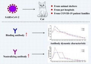
In this study, 39 banked sera collected from cats before the outbreak (March to May 2019) and 102 samples collected from cats in animal shelters and veterinary clinics in Wuhan from January to March 2020 (i.e. during the COVID-19 outbreak) were screened by indirect enzyme linked immunosorbent assay (ELISA) for antibody reactivity against recombinant RBD of SARS-CoV-2 spike protein.
15 (14.7%) of the samples collected from January to March 2020 were positive for RBD-based ELISA and 11 of these then showed neutralizing antibodies against SARS-CoV-2 when tested by virus neutralization tests.
The cats with the highest titres belonged to people with confirmed COVID-19. While it may not be surprising that cats in contact with owners infected with COVID-19 can become infected, it is interesting that a number of other cats also mounted some degree of immune response.
It was also noted that both type I and II feline infectious peritonitis virus (FIPV) hyperimmune sera showed no cross-reactivity withSARS-CoV-2 RBD protein.
In addition, the researchers continuously monitored serum antibody dynamics of two positive cats every 10 days over 130 days. The authors reported that serum antibodies reached the peak at 10 days after first sampling and declined to the limit of detection within 110 days.
While this study provides some preliminary evidence that cats can become infected, and mount an immune response raising an antibody response to SARS-CoV-2, it did not give any information on whether the cats are able to pass on the virus.
Image reproduced under Creative Commons CC BY license from A serological survey of SARS-CoV-2 in cat in Wuhan. Emerging Microbes & Infections, 9 (1), p 2013
Reference for pre print:
Zhang, Q. et al. (2020) SARS-CoV-2 neutralizing serum antibodies in cats: a serological investigation bioRxiv https://doi.org/10.1101/2020.04.01.021196
- Temmam, S. et al. (2020) Absence of SARS-CoV-2 infection in cats and dogs in close contact with a cluster of COVID-19 patients in a veterinary campus. One Health, 10, no. 100164. https://doi.org/10.1016/j.onehlt.2020.100164
This study, from the Institut Pasteur, reports on the testing of 9 cats and 12 dogs living in close contact with their owners, belonging to a group of 20 veterinary students in which two students tested positive for COVID-19 and several others (n = 11/18) showed clinical signs (fever, cough, anosmia, etc.) consistent with COVID-19 infection, between 25th February and 18th March 2020. Although a few pets were reported to have presented clinical signs indicative of a coronavirus infection; blood samples collected on 25th March and nasal and rectal swabs collected daily for 1 week, starting from the day of blood sampling, all tested negative for virus (PCR) and antibodies (immunoprecipitation).
While this study provides preliminary evidence that animals living with their owners do not become infected, it is important to note that this is a small study and it is possible that the timing of the samples could have missed transient contamination.
Ferrets/minks
- Santman-Berends, I.M.G.A. et al. (2024) Effectiveness of passive and active surveillance for early detection of SARS-CoV-2 in mink during the 2020 outbreak in the Netherlands. Transboundary and Emerging Diseases, no. 4793475. https://doi.org/10.1155/2024/4793475
- Himsworth, C. G. et al. (2023) A comparison of sampling and testing approaches for the surveillance of SARS-CoV-2 in farmed American mink. Journal of Veterinary Diagnostic Investigation, 35 (5), pp. 528-534. https://doi.org/10.1177/10406387231183685
- Žigaitė, S. et al. (2023) Evaluation of SARS-CoV-2 passive surveillance in Lithuanian mink farms, 2020–2021. Frontiers in Veterinary Science, 10. https://doi.org/10.3389/fvets.2023.1181826
- Kaczorek-Łukowska, E. et al. (2023) No indication for SARS-CoV-2 transmission to pet ferrets, in five cities in Poland, 2021 – antibody testing among ferrets living with owners infected with SARS-CoV-2 or free of infection. Acta Veterinaria Scandinavica, 65, no. 9. https://doi.org/10.1186/s13028-023-00672-3
- Sikkema, R. S. et al. (2022) Risks of SARS-CoV-2 transmission between free-ranging animals and captive mink in the Netherlands. Transboundary and Emerging Diseaseshttps://doi.org/10.1111/tbed.14686
- Davoust, B. et al. (2022) Evidence of antibodies against SARS‐CoV‐2 in wild mustelids from Brittany (France). Transboundary and Emerging Diseaseshttps://doi.org/10.1111/tbed.14663
- Cai, H.Y. and Cai, A. (2021) SARS-CoV2 spike protein gene variants with N501T and G142D mutation–dominated infections in mink in the United States. Journal of Veterinary Diagnostic Investigation, 33 (5) pp. 939-942. https://doi.org/10.1177/10406387211023481
Report on genetic characteristics of the U.S. and Canadian mink–derived SARS-CoV2 sequences. The study reports that novel SARS-CoV2 variants are most likely to have evolved during human infection and were then transmitted to mink populations in the United States.
- Rabalski, L. et al (2021) Severe acute respiratory syndrome coronavirus 2 in farmed mink (Neovison vison), Poland. Emerging Infectious Diseases, 27 (9), p. 2333-2339. https://doi.org/10.3201/eid2709.210286
Report of a COVID outbreak in farmed mink in Northern Poland reporting test results and genetic sequencing.
- van Aart, A.E. et al (2021) SARS‐CoV‐2 infection in cats and dogs in infected mink farms. Transboundary and Emerging Diseases. https://doi.org/10.1111/tbed.14173
This short communication reports on the prevalence of SARS-CoV-2-positive cats and dogs from ten infected mink farms in the Netherlands, and their possible role in transmission of the virus.
Throat and rectal swabs of 101 cats (12 domestic and 89 feral cats) and 13 dogs were tested for SARS-CoV-2 using PCR. Eleven cats (18%) and two dogs (15%) tested serologically positive. Three feral cats (3%) and one dog (8%) tested PCR-positive.
The authors conclude that as only feral cats were infected it is most likely that infections in cats were initiated by mink, not by humans. Whether both dogs were infected by mink or humans remains inconclusive.
- Gortazar, C. et al (2021) Natural SARS-CoV-2 infection in kept ferrets, Spain. Emerging Infectious Diseases, 27 (7), pp. 1994-1996. https://doi.org/10.3201/eid2707.210096
This research letter reports on SARS-CoV-2 infection in 71 ferrets belonging to 7 owners; the ferrets were used as working animals for rabbit hunting in Ciudad Real Province, central Spain. SARS-CoV-2 RNA was found in swab samples (1 rectal and 5 nasal) from 6 (8.4%) of the 71 ferrets from 4 of the 7 groups of ferrets investigated.
The authors conclude that natural SARS-CoV-2 infection in kept ferrets does occur in circumstances of high viral circulation in the human population. However, the high cycle thresholds observed and the lack of virus-positive ferrets at resampling suggest that small ferret populations are less able to maintain prolonged virus circulation than large, farmed mink populations.
- Shriner, S.A. et al (2021) SARS-CoV-2 exposure in escaped mink, Utah, USA. Emerging Infectious Diseases, 27 (3), pp. 988-990. https://dx.doi.org/10.3201%2Feid2703.204444
This paper reports on a wildlife epidemiologic investigation of mammals captured on or near properties in Utah, USA, where outbreaks of severe acute respiratory syndrome coronavirus 2 (SARS-CoV-2) infection occurred in farmed mink.
Free-roaming mammals were captured between August 22–30, 2020, by using Sherman (rodents) and Tomahawk (mesocarnivores) traps placed outside barns and barrier fences on outbreak premises and public lands within a 3.5-km buffer zone. Sample collection included oral, nasal (washes for mice), and rectal swabs as well as tissue and blood samples.
102 mammals were captured (78 rodents and 24 mesocarnivores). Rodents captured consisted of three species of mice and three rock squirrels. Mesocarnivore captures consisted of 11 presumed escaped American mink, two presumed wild American mink, five raccoons and six striped skunks. Presumed escaped mink were closely associated with barns and designated as domestic escapees on the basis of location, behaviour, and appearance.
Wild mink were identified by brown coat colour and smaller size compared with farmed mink.
All escaped mink and rodents, except for four deer mice and one rock squirrel, were caught on farm premises. All raccoons, the two presumed wild mink, and all but one striped skunk were captured off-property but within the buffer zone.
Serum samples from the 11 mink escapees tested positive for SARS-CoV-2 antibodies by virus neutralization, and three also had viral RNA detected by rRT-PCR from nasal swabs and one from lung tissue. A rectal swab specimen from a house mouse had a high Ct detection by rRT-PCR but was negative for SARS-CoV-2 antibodies.
No other animal had a detectable antibody response.
Although the authors did not find evidence for SARS-CoV-2 establishment in wildlife, they note that the discovery of escaped mink with the opportunity to disperse and interact with susceptible wildlife, such as wild mink or deer mice, is concerning and recommend heightened biosecurity to help prevent accidental releases of infected animals or spillover of SARS-CoV-2 from susceptible species to native wildlife.
- Hammer, A.S. et al (2021) SARS-CoV-2 transmission between mink (Neovison vison) and humans, Denmark. Emerging infectious diseases, 27 (2), pp. 547-551. https://dx.doi.org/10.3201/eid2702.203794
This paper reports on the epidemiological investigation into SARS-CoV-2 infection at three mink farms in the Northern Jutland region of Denmark, to analyse the transmission of virus in mink and the local human community.
Swab samples (blood and throat, nasal, and faecal swabs) were collected from adult mink and kits from 3 different mink farms. Air and feed samples were also collected. Samples were assessed for viral RNA by quantitative reverse transcription PCR (qRT-PCR) and SARS-CoV-2 Ab ELISA.
At initial sampling, seroprevalence was high on farm 1 (>95%) and farm 3 (66%) but, only 3% on farm 2. However, at follow up sampling seroprevalence on farm 2 had increased to >95%.
The authors note that despite the high level of virus detected in the mink there was little clinical disease or increase in death rate, making it difficult to detect the spread of infection; thus, mink farms could represent a serious, unrecognized animal reservoir for SARS-CoV-2.
Air samples from farm 1 tested negative. However, on farms 2 and 3, multiple samples collected from exhaled air from mink or within 1 m of the cages were positive. None of the air samples collected outside the houses were positive. Feed samples collected at each farm tested negative.
SARS-CoV-2–positive samples were then sequenced. The viruses found on farms 1–3 were very similar and these sequences and those from humans linked to the infected farms grouped within the European 20B clade of the global SARS-CoV-2 tree.
The authors conclude that a likely scenario for the spread of infection in mink in Denmark is that the index human case-patient introduced infection into farm 1, where a mutation occurred that could be linked to subsequent human cases. It seems that the variant viruses on farm 1 spread to >1 human and were then transmitted, presumably by human–human contact, to other people and to farms 2 and 3.
- Sawatzki, K. et al. (2020) Ferrets not infected by SARS-CoV-2 in a high-exposure domestic setting. bioRxiv. https://doi.org/10.1101/2020.08.21.254995
This study reports on the results of a “natural experiment” where 29 ferrets in one home had prolonged, direct contact and constant environmental exposure to two humans with symptomatic COVID-19. The authors observed no evidence of SARS-CoV-2 transmission from humans to ferrets based on RT-PCR and ELISA.
This research was carried out as part of the Coronavirus Epidemiological Response and Surveillance (CoVERS) study, set up at Tufts University to investigate the potential for human-to animal spill over and onward transmission in domestic, farm and wildlife species.
A household with 29 free-roaming ferrets cared for by two adults was enrolled as part of the CoVERS study. Individual 1 experienced fever and fatigue from 25 March-6 April and individual 2 experienced a sore throat, anosmia, migraine and fatigue from 28 March-13 April. Individual 2 tested positive for SARS-CoV-2/COVID-19 infection by nasopharyngeal swab and RT-PCR on 1 April. Individual 1 was a probable positive due to the timing and symptoms but was not tested. Neither person was hospitalised, and both cared for the ferrets during the entirety of their disease courses.
A two-week, in-home sample collection scheme was designed to begin during the household quarantine period, the ferrets were free to move in all spaces of the home during this period and were handled as usual, including regular petting, feeding and grooming. A home sampling kit sent to the participants including material to safely collect and store ferret oral swabs. One participant had significant animal handling experience and performed all sample collection to standardise sampling procedures. Thirty oral swabs were collected and held in viral transport media in the participants’ freezer until the end of the study period. Frozen samples were directly transferred to a lab member and processed.
Oral swabs were collected from all ferrets in the home over a two-week period, beginning 10 April, concurrent with symptomatic disease in individual 2. One ferret (3) was sampled twice. Two 7-year-old ferrets (12 and 16) died during the study period, one by euthanasia due to chronic disease, the other cause is unknown. Thirty samples from 29 ferret oral swabs were tested by semi-quantitative real time RT-PCR and ELISA.
All samples were confirmed to have viable RNA (by a preliminary screen for constitutively expressed ß-actin) but results of semi-quantitative real time RT-PCR and ELISA were below the limit of detection and determined to be negative for active or recent infection by SARS-CoV-2.
As ferrets have been shown to be susceptible to infection and onward transmission in experimental laboratory infections the researchers undertook further analysis to better understand this discrepancy in experimental and natural infection in ferrets.
They compared SARS-CoV-2 sequences from natural and experimental mustelid infections and identified two surface glycoprotein (Spike) mutations. Evidence found that while ACE2 provides a weak host barrier, one mutation only seen in ferrets is located in the novel S1/S2 cleavage site and is computationally predicted to decrease furin activity. They conclude that the data support that host factors interacting with the novel S1/S2 cleavage site may be a barrier in ferret SARS-CoV-2 susceptibility and that domestic ferrets are at low risk of natural infection from currently circulating SARS-CoV-2. This may be overcome in laboratory settings using concentrated viral inoculum, but the effects of ferret host-adaptations require additional investigation.
- Munnink, B.B.O. et al. (2020) Jumping back and forth: anthropozoonotic and zoonotic transmission of SARS-CoV-2 on mink farms. bioRxiv, 2020.09.01.277152 https://doi.org/10.1101/2020.09.01.277152
This paper describes an in-depth investigation of SARS-CoV-2 outbreaks on 16 mink farms, in the Netherlands, and infection in the people living or working on these farms, combining epidemiological information, surveillance data and whole genome sequencing (WGS).
97 individuals were tested by either serological assays and/or RT-PCR. 43 out of 88 (49%) of upper-respiratory tract samples tested positive by RT-PCR while 38 out of 75 (51%) of serum samples tested positive for SARS CoV-2 specific antibodies. In total, 66 of 97 (67%) of the people tested had evidence for SARS CoV-2 infection. To maintain anonymity the farms were grouped into geographic areas for analysis.
The whole genome sequences generated from mink farms and from mink farm employees were compared with the national database consisting of around 1,775 WGS. In addition, to discriminate between locally acquired infections and mink farm related SARS-CoV-2 infection, and to determine the potential risk for people living close to mink farms, WGS was also performed on 34 SARS-CoV-2 positive samples from individuals who live in the same four-digit postal code area as the first four mink farms. These local sequences reflected the general diversity seen in the Netherlands and were not related to the clusters of mink sequences found on the mink farms, giving no indication of spill-over to people living in close proximity to mink farms.
After the detection of SARS-CoV-2 on mink farms, 68% of the tested farm workers and/or relatives or contacts were shown to be infected with SARS CoV-2, indicating that contact with SARS-CoV-2 infected mink is a risk factor for contracting COVID-19.
A high diversity in the sequences from some mink farms was observed which the authors considered was most likely explained by many generations of infected animals before an increase in mortality was observed. They note that mink farms have large populations of animals which could lead to very efficient virus transmission and that the virus might replicate more efficiently in mink or might have acquired mutations which makes the virus more virulent.
A mutation in the spike protein (D614G), that has been shown to result in an increased virulence in vitro, was present in farm clusters A, C and E, but no obvious differences in clinical presentation, disease severity, or rate of transmission to humans was observed.
The authors conclude that the virus was initially introduced from humans and has evolved, most likely reflecting widespread circulation among mink in the beginning of the infection period, several weeks prior to detection.
Please note this paper has been published as a preprint on bioRxiv and has not been subject to peer review.
Bats
- Common, S.M. et al (2021) The risk from SARS‐CoV‐2 to bat species in England and mitigation options for conservation field workers. Transboundary and Emerging Diseases. https://doi.org/10.1111/tbed.14035
This paper reports on a qualitative disease risk analysis (DRA), undertaken to assess the risk of disease from SARS‐CoV‐2 to free‐living bats from fieldworkers carrying out bat conservation interventions and development activities in England. The probability of disease occurring and the magnitude of the possible consequences to bat populations were assessed and mitigation methods proposed.
The disease risk assessment was carried out by staff from The Institute of Zoology and Natural England, according to the method described by the OIE. Assessment was undertaken of the biological pathways that might permit bats to be exposed and infected with SARS‐CoV‐2, as well as the probability of exposure and infection occurring.
The probability of exposure of bats to SARS‐CoV‐2 through fieldwork activities was estimated to range from negligible to high, depending on the proximity between bats and people during the activity. The likelihood of infection after exposure was estimated to be high and the probability of dissemination of the virus through bat populations medium.
There is uncertainty in the pathogenicity of SARS‐CoV‐2 in bats, with the authors reporting that SARS‐CoV‐2 has been demonstrated experimentally to infect one species of bat, but another has been shown to be experimentally resistant. Therefore, although the likelihood of clinical disease occurring in infected bats was assessed as low there is some uncertainty in this risk estimation. The authors note that the disease risk analysis should be updated as information on the epidemiology of SARS‐CoV‐2 and related viruses in bats improves.
The authors conclude that the probability of infection can be effectively reduced if fieldworkers follow routine government guidance, and minimum precautions have been set out in advice provided by DEFRA to Natural England and in addition follow strict biosecurity measures when contacting bats or possible fomites which may expose bats to the virus, including the use of disposable gloves, cloth face coverings, effective hand cleansing and appropriate disinfecting of equipment.
Guidance
Natural England (2020). COVID‐19 and interacting with wildlife for the purposes surveying and mitigation works [online] Available from: https://www.gov.uk/guidance/coronavirus-covid-19-surveying-and-mitigation-works-affecting-wildlife [Accessed 22 March 2021]
Botto Nunez, G. et al (2020) IUCN SSC Bat Specialist Group (BSG) recommended Strategy for researchers to reduce the risk of transmission of SARS-CoV-2 from humans to bats [online] Available from: https://www.iucnbsg.org/uploads/6/5/0/9/6509077/map_recommendations_for_researchers_v._1.0_final.pdf [Accessed 23 September 2025]
Deer
- Tarbuck, N.N. et al. (2025) Persistence of SARS-CoV-2 Alpha variant in white-tailed deer, Ohio, USA. Emerging Infectious Diseases, 31 (7), pp. 1319-1329. https://doi.org/10.3201/eid3107.241922
- Rosenblatt, E.G. et al. (2025) Fomites could determine severity of SARS‐CoV‐2 outbreaks in low‐density white‐tailed deer (Odocoileus virginianus) populations. Transboundary and Emerging Diseases. https://doi.org/10.1155/tbed/1352911
- Purves, K. et al. (2024) SARS-CoV-2 seropositivity in urban population of wild fallow deer, Dublin, Ireland, 2020-2022. Emerging Infectious Diseases, 30 (8), pp. 1609-1620. https://doi.org/10.3201/eid3008.231056
- McBride, D.S. et al. (2023) Accelerated evolution of SARS-CoV-2 in free-ranging white-tailed deer. Nature Communications, 14, no. 5105. https://doi.org/10.1038/s41467-023-40706-y
- Khalafalla, A. I. (2023) SARS-CoV-2 Neutralizing Antibodies in Free-Ranging Fallow Deer (Dama dama) and Red Deer (Cervus elaphus) in Suburban and Rural Areas in Spain. Transboundary and Emerging Diseases, no. 3324790. https://doi.org/10.1155/2023/3324790
- OIE Statement on monitoring white-tailed deer for SARS-CoV-2. World Organization for Animal Health [online] Available from: https://www.oie.int/en/oie-statement-on-monitoring-white-tailed-deer-for-sars-cov-2/ [accessed 1 February 2022]
Published 1 February 2022
- USDA Animal and Plant Health Inspection Service (2021) Surveillance Data Shows White-Tailed Deer Exposed to SARS-CoV-2 [online] Available from: https://www.aphis.usda.gov/aphis/newsroom/stakeholder-info/stakeholder-messages/wildlife-damage-news/deer-sars [Accessed 6 August 2021]
The U.S. Department of Agriculture has released a report of a surveillance study that analysed serum samples from free ranging white-tailed deer for antibodies to SARS-CoV-2.
Samples were collected in 4 states (Illinois, Michigan, New York, and Pennsylvania) between January 2020 and January 2021. None of the deer populations surveyed showed signs of clinical illness associated with SARS-CoV-2, and evidence of viral shedding was not undertaken. However, antibodies to SARS-CoV-2 were detected in 33% of the 481 samples, indicating exposure. Concerns that the test, which has not yet been validated in deer, may have been cross reacting with anther virus, were addressed by retesting the samples using a test specific to SARS-CoV-2 and testing archived samples from before the pandemic.
The report states that the finding that wild white-tailed deer have been exposed to SARS-CoV-2 is not unexpected given that white-tailed deer are susceptible to the virus, are abundant in the United States, often come into close contact with people, and that, more than 114 million Americans are estimated to have been infected with COVID-19, according to the U.S. Centers for Disease Control and Prevention. However, this level of exposure raises significant questions about this risk of SARS -CoV-2 becoming established in wildlife, and further research is urgently needed.
Equine
- Pusterla, N., Lawton, K. and Barnum, S. (2023) Investigation of the seroprevalence to equine coronavirus and SARS-CoV-2 in healthy adult horses recently imported to the United States. Veterinary Quarterly. https://doi.org/10.1080/01652176.2023.2288876
Wildlife and zoo
- Wilson-Henjum, G. et al. (2025) Community-scale surveillance of SARS-CoV-2 and influenza A viruses in wild mammals, United States, 2022–2023. Emerging Infectious Diseases, 31 (8), pp. 1625-1629. https://doi.org/10.3201/eid3108.241671
- Didkowska, A. et. al. (2025) Presence of anti-SARS-CoV-2 antibodies in European bison (Bison bonasus) in Poland, 2019–2023. BMC Veterinary Research, 21, no. 120. https://doi.org/10.1186/s12917-025-04593-3
- Loy, D.S. et al. (2025) SARS-CoV-2 surveillance and detection in wild, captive, and domesticated animals in Nebraska: 2021–2023. Frontiers in Veterinary Science, 11. https://doi.org/10.3389/fvets.2024.1496207
- Domanska-Blicharz, K., Lisowska, A., Opolska, J., Ruszkowski, J., Gogulski, M., Pomorska-Mól, M. (2024) Whole genome characteristics of hedgehog coronaviruses from Poland and analysis of the evolution of the Spike protein for its interspecies transmission potential. BMC Veterinary Research. https://doi.org/10.1186/s12917-024-04277-4
- Cano-Terriza, D. et al. (2024) SARS-CoV-2 in captive nonhuman primates, Spain, 2020-2023. Emerging Infectious Diseases, 30 (6), pp. 1253-1257. https://doi.org/10.3201/eid3006.231247
- Takemura, T. et al. (2024) SARS-CoV-2 Infection in Beaver Farm, Mongolia, 2021. Emerging Infectious Diseases, 30 (2), pp. 391-394. https://doi.org/10.3201/eid3002.231318
- Ehrlich, M. et al. (2023) Lack of SARS-CoV-2 Viral RNA Detection among a Convenience Sampling of Ohio Wildlife, Companion, and Agricultural Animals, 2020–2021. Animals, 13 (16), no. 2554. https://doi.org/10.3390/ani13162554
- Colombo, V.C. et al. (2022) SARS‐CoV‐2 surveillance in Norway rats (Rattus norvegicus) from Antwerp sewer system, Belgium. Transboundary and Emerging Diseases, 69 (5), pp. 3016-3021. https://doi.org/10.1111/tbed.14219.
- Wang, Y. et al. (2022) SARS-CoV-2 exposure in Norway rats (Rattus norvegicus) from New York City. bioRxiv [Preprint]. https://doi.org/10.1101/2022.11.18.517156.
Reviews
- Awada, L. et al. (2024) Facing SARS-CoV-2 emergence on the animal health perspective: The role of the World Organisation for Animal Health in preparedness and official reporting of disease occurrence. Zoonoses and Public Health. https://doi.org/10.1111/zph.13133
- Nederlof, R.A., de la Garza, M.A. and Bakker, J. (2024) Perspectives on SARS-CoV-2 Cases in Zoological Institutions. Veterinary Sciences, 11 (2), no. 78. https://doi.org/10.3390/vetsci11020078
- Laconi, A. et al. (2023) SARS-CoV-2 and companion animals: Sources of information and communication campaign during the COVID-19 pandemic in Italy. Veterinary Sciences, 10 (7), no. 426. https://doi.org/10.3390/vetsci10070426
- Joffrin, L. et al. (2023) SARS-CoV-2 Surveillance between 2020 and 2021 of all mammalian species in two Flemish zoos (Antwerp Zoo and Planckendael Zoo). Veterinary Sciences, 10 (6), no. 382. https://doi.org/10.3390/vetsci10060382
- Reggiani, A., Rugna, G. and Bonilauri, P. (2022) SARS-CoV-2 and animals, a long story that doesn’t have to end now: What we need to learn from the emergence of the Omicron variant. Frontiers in Veterinary Science, 9. https://doi.org/10.3389/fvets.2022.1085613.
- Hobbs, E.C. and Reid, T.J. (2020). Animals and SARS‐CoV‐2: Species susceptibility and viral transmission in experimental and natural conditions, and the potential implications for community transmission. Transboundary and Emerging Diseases. https://doi.org/10.1111/tbed.13885
This paper, first published on 22 October 2020, presents the findings of a scoping literature review conducted to collect, evaluate and present the available research evidence regarding SARS‐CoV‐2 infections in animals. The authors include both experimental studies and reports of natural infection and conclude that “Most animals are presumed to have been infected by close contact with COVID‐19 patients. In domestic settings, viral transmission is self‐limiting; however, in high‐density animal environments, there can be sustained between‐animal transmission”.
- de Morais, H.A. (2020) Natural infection by SARS-CoV-2 in companion animals: a review of case reports and current evidence of their role in the epidemiology of COVID-19. Frontiers in Veterinary Science, 7, p.823. https://doi.org/10.3389/fvets.2020.591216
This paper provides a review of reported cases of animals naturally infected with SARS-CoV-2, particularly companion pets, with the aim of shedding light on the role of these animals in the epidemiology of COVID-19. It includes a brief overview of coronaviruses and information on the similarity between the ACE2 protein in various animals and humans, which may have implications for susceptibility to infection with SARS-CoV-2.
- Mathavarajah, S. (2020) Pandemic danger to the deep: the risk of marine mammals contracting SARS-CoV-2 from wastewater. Science of The Total Environment, p.143346. https://doi.org/10.1016/j.scitotenv.2020.143346
This paper reports on an analysis of ACE2 receptors using annotated genomes of marine mammals from the 4 major groups (36 species) in order to generate an index of susceptibility for marine mammals to SARS-CoV-2.
To distinguish between the susceptible and non-susceptible species, the authors used the human ACE2 (high susceptibility), feline ACE2 (medium susceptibility; lower affinity but still susceptible) and dog ACE2 (not susceptible) as reference points. Using this method, they identified that many species of whale, dolphin, and seal, as well as otters, are predicted to be highly susceptible to infection by the SARS-CoV-2 virus.
The authors then looked at wastewater management in certain Alaskan localities and concluded that this may not be sufficient for preventing waterborne exposure of nearby marine mammals to the virus.
They concluded that while the risk to marine mammals is likely very low, especially in terms of creating a sustained problem , since some marine mammal populations are highly threatened, an outbreak localized to an individual pod or population could still have significant consequences.
- Alexander, M.R. (2020) Predicting susceptibility to SARS‐CoV‐2 infection based on structural differences in ACE2 across species. The FASEB Journal. 34 (2) pp. 15946-15960 https://doi.org/10.1096/fj.202001808R
This paper reports on potential species differences in susceptibility to SARS-CoV-2 using multiple in-depth structural analyses to identify key ACE2 amino acid positions (including 30, 83, 90, 322, and 354) and use these differences to develop a susceptibility score.
The authors conclude that SARS-CoV-2 is nearly optimal for binding ACE2 of humans compared to other animals, which may underlie the highly contagious transmissibility of this virus among humans.
While this study does not give us information about what is happening in terms of infection these species it may provide information to direct future surveillance and research.
- OiE (2020) Infection with SARS-COV2 in animals [online] Available at https://www.oie.int/fileadmin/Home/MM/A_Factsheet_SARS-CoV-2__1_.pdf [Accessed 17 July 2020]
The OiE have produced a technical factsheet which provides a brief summary of the current (June 2020) knowledge about SARS-CoV-2 infection in animals. It includes sections on aetiology, epidemiology, diagnosis, and methods of prevention and control. It also includes this table summarising current knowledge about infection in animals.
| SPECIES | TYPE OF INFECTION | SUSCEPTIBILITY | CLINICAL SIGNS | TRANSMISSION |
|---|---|---|---|---|
| Pigs | Experimental | None | No | No |
| Poultry (chickens, ducks and turkeys) | Experimental | None | No | No |
| Dogs | Natural and experimental | Low | No (possible some cases) | No |
| Cats (domestic) | Natural and experimental | High | Yes (none to very mild in some cases) | Yes, between cats |
| Tigers and lions | Natural | High | Yes | Yes, between animals |
| Ferrets | Experimental | High | No (mild in some cases) | Yes, between ferrets |
| Minks (American minks, Neovison vison) | Natural | High | Yes | Yes, between minks and suggested from minks to humans |
| Egyptian fruit bats (Rousettus aegyptiacus) | Experimental | High | No | Yes, between fruit bats |
| Golden Syrian Hamsters | Experimental | High | Yes (none to very mild in some cases) | Yes, between hamsters |
| Macaques (Macaca fascicularis and Macaca mulatta) | Experimental | High | Yes | Yes |
- Decaro, N. et al. (2020) COVID-19 from veterinary medicine and one health perspectives: What animal coronaviruses have taught us. Research in Veterinary Science, 131, pp. 21-23. https://doi.org/10.1016/j.rvsc.2020.04.009
This review article provides a brief overview of animal coronaviruses and the veterinary experience of dealing with them. The authors note that there is extensive knowledge in veterinary medicine about animal coronaviruses, their evolution and pathobiology. They provide brief details of the major veterinary coronaviruses, such as Infectious Bronchitis Virus (IBV) of poultry and Feline Infectious Peritonitis Virus (FIPV) which have been known since the early 1900s, and provide examples on how coronaviruses can evolve, changing their tissue tropism and virulence. The authors use as an example the
Transmissible Gastro-Enteritis Virus of pigs (TGEV), which likely originated from the closely related canine coronavirus (CCoV), and in turn gave rise to the less virulent Porcine Respiratory Coronavirus (PRCoV).
The authors note that while animal models may be useful in developing human SARS-CoV-2 vaccines, there may also be lessons to be learned from experience using veterinary vaccines such as those for IBV and CCoV. In these cases, parentally administered vaccines against respiratory coronaviruses have been found to reduce the severity of respiratory signs, but not give full protection against respiratory infection or virulent virus, noting that prevention of infection may be more dependent on mucosal immunity.
Also discussed is the issue of antibody-dependent enhancement, which was found to cause a more severe disease in cats immunised against Feline Infectious Peritonitis Virus (FIPV) than in control cats.
A more positive lesson from the management of FIP relates to recent attempts to control FIP using two promising antiviral classes, namely protease inhibitors and nucleoside analogues, such as GS-441524, which is similar to the adenosine nucleoside monophosphate prodrug GS-5734; GS-5734 is the active molecule of Remdesivir.
The authors conclude that given the long-term experience gained with animal coronaviruses, veterinary medicine could help to forge a better understanding of the origin and spread of SARS-CoV-2 and guide future research in human medicine towards the development of immunogenic and safe vaccines and effective antiviral drug.
- O’Connor, A.M, Totton, S.C. and Sargeant, J.M. (2020) SYREAF [Systematic Reviews for Animals and Food]: a rapid review of evidence of infection of pets and livestock with human-associated coronavirus diseases, SARS, MERS, and COVID-19, and evidence of the fomite potential of pets and livestock. https://www.cabidigitallibrary.org/doi/full/10.5555/20203180496
The review looked at the evidence available to answer two questions
Question 1: “What is the evidence that domestic animals (cats, ferrets, dogs, swine, cattle, sheep, goats, poultry, horses) can be infected with, or shed, the human-associated coronaviruses SARS-CoV, MERS-CoV, and SARS-CoV-2, which are associated with the diseases, SARS, MERS, and COVID-19, respectively?”
Question 2: “What is the evidence that domestic animals (cats, ferrets, dogs, swine, cattle, sheep, goats, poultry, horses) can act as a fomite for the human-associated coronaviruses SARS-CoV, MERS- CoV, and SARS-CoV-2, which are associated with the diseases, SARS, MERS, and COVID-19, respectively?
The review includes case studies and early experimental and epidemiological studies as well as studies of related coronaviruses (SARS and MERS).
They concluded that from the evidence reviewed (to 29th April 2020):
- All reported cases where the SARS-CoV-2 virus has been detected in cats or dogs have been living in close quarters with SARS-CoV-2 infected owners. Two cats and two dogs have been reported to have SARS-CoV-2 virus detected by PCR.
- Fifteen of 102 cats in Wuhan China have been reported as positive for antibodies to SARS-CoV-2.
- No cases of cat- or dog-to-human transmission have been reported. Two SARS-CoV-2 seeder challenge studies have reported no transmission to contact pigs, chickens or ducks.
- In a survey of 12 dogs and nine cats living in close contact with 20 veterinary students (including 2 confirmed and 11 suspected COVID-19 patients) all animals were negative for antibodies and PCR to SARS-CoV-2.
- One study showed that SARS-CoV can survive on room temperature pig skin for >24h. No other studies were found that evaluated fur, hair, skin, feathers, or hides as a source of transmission from domestic animals for SARS, MERS, or SARS-CoV-2.
The authors acknowledge that there are many questions unanswered and state that they see this as a living review which will be updated as new evidence becomes available.
It should be noted that while this is described as a “systematic review”, this review of the published literature was undertaken rapidly in response to the current pandemic and has not been peer-reviewed.
Impact of the pandemic
There are now a number of articles and surveys being published on the impact of the COVID pandemic on veterinary practice, animal ownership and animal welfare. Links, and a brief description of each of these papers, are provided below:
- Humer, E. et al. (2024) Veterinary medicine under COVID-19: a mixed-methods analysis of student and practitioner experiences in Austria. Frontiers in Veterinary Science, 11. https://doi.org/10.3389/fvets.2024.1460269
- Yi, J. et al. (2024) Student and clinical educator perceptions of the impacts of COVID-19 on final-year veterinary clinical training in a distributed learning model. Journal of Veterinary Medical Education, 51 (4), pp. 461-472. https://doi.org/10.3138/jvme-2023-0004
- Langebæk, R. et L. (2024) A collaborative response to the COVID-19 challenge: Developing an international platform for sharing e-learning materials for veterinary education. Journal of Veterinary Medical Education, 51 (4), pp. 42-430. https://doi.org/10.3138/jvme-2023-0039
- Germann, J.A. et al. (2024) Biosecurity perceptions among Ontario horse owners during the COVID-19 pandemic. Equine Veterinary Journal. https://doi.org/10.1111/evj.14115
- King, E.K. et al. (2024) Longitudinal patterns of companion animals in families with children during the COVID-19 pandemic: Findings from the Adolescent Brain Cognitive Development (ABCD) Study®. Frontiers in Veterinary Science, 11. https://doi.org/10.3389/fvets.2024.1364718
- Luethy, D. et al. (2023) Cross-sectional study of physical activity, dietary habits, and mental health of veterinary students after lifting of COVID-19 pandemic measures. PLOS ONE, 18(9), no. e0291590. https://doi.org/10.1371/journal.pone.0291590
- Burzette, R.G. (2023) An exploratory study of the impact of COVID-19 pandemic disruptions on veterinary medical education. Journal of Veterinary Medical Education. https://doi.org/10.3138/jvme-2023-0049
- Manning, P. (2023) The effect of COVID-19 lockdown restrictions on self-directed behaviour, activity budgets, movement patterns, and spatial use in semi-captive African elephants (Loxodonta africana). Applied Animal Behaviour Science, 266. https://doi.org/10.1016/j.applanim.2023.106007
Veterinary practice
- Elane, G.L., Blikslager, A.T. and Mair, T.S. (2024) Trends in the management of horses referred for colic evaluation preceding and during the COVID-19 pandemic (2013–2023). Equine Veterinary Education. https://doi.org/10.1111/eve.14038
- Kanwischer, M., Tipold, A. and Schaper, E. (2024) Veterinary teaching in COVID-19 times: perspectives of university teaching staff. Frontiers in Veterinary Science, 11. https://doi.org/10.3389/fvets.2024.1386978
- Schroeder, C.A. (2024) Perceived impact of the COVID-19 pandemic on residency training in American College of Veterinary Anesthesia and Analgesia programs in North America: a quantitative survey. Veterinary Anaesthesia and Analgesia. https://doi.org/10.1016/j.vaa.2024.06.007
- Nichelason, A. and Genovese, J. (2023) The COVID-19 pandemic negatively impacted veterinary client satisfaction and loyalty. Journal of the American Veterinary Medical Association. https://doi.org/10.2460/javma.23.06.0351
- Darby, B.J. et al. (2023) Veterinarians show resilience during COVID-19: challenges faced and successful coping strategies. Journal of the American Veterinary Medical Association.https://doi.org/10.2460/javma.22.12.0584
- Russon, J.M. et al. (2023) Career stage differences in mental health symptom burden and help seeking among veterinarians during COVID-19. Journal of the American Veterinary Medical Association. https://doi.org/10.2460/javma.22.12.0583
- Dubin, R.J. et al (2021) Veterinarians’ perceptions of COVID-19 pandemic–related influences on veterinary telehealth and on pet owners’ attitudes toward cats and dogs. Journal of the American Veterinary Medical Association, 259 (10), pp. 1140-1147. https://doi.org/10.2460/javma.21.04.0203
- Smith, S.M. et al (2022) Opportunities for expanding access to veterinary care: lessons from COVID-19. Frontiers in Veterinary Science, 9, no. 804794. https://doi.org/10.3389/fvets.2022.804794
- Hoffman, C.L., Thibault, M. and Hong, J.(2021) Characterizing pet acquisition and retention during the COVID-19 pandemic. Frontiers in Veterinary Science, 8, p. 1375. https://doi.org/10.3389/fvets.2021.781403
- Limper, C.B., Hinckley-Boltax, A.L. and Cazer, C.L. (2021) Brief Research Report: Veterinary student perspective on COVID-19 and veterinary medicine. Frontiers in Veterinary Science, 8, p. 1213. https://doi.org/10.3389/fvets.2021.723890
- McKee, H. et al (2021) High psychosocial work demands, decreased well-being, and perceived well-being needs within veterinary academia during the COVID- 19 pandemic. Frontiers in Veterinary Science, 8, p. 1200. https://doi.org/10.3389/fvets.2021.746716
- Morris, A., Wu, H. and Morales, C. (2021) Barriers to care in veterinary services: lessons learned from low-income pet guardians’ experiences at private clinics and hospitals during COVID-19. Frontiers in Veterinary Science, 8, p. 1227. https://doi.org/10.3389/fvets.2021.764753
- Mureşan, A.N., Morariu, S., Baisan, R.A., Costea, R. and Mureşan, C., 2021. The impact of COVID-19 pandemic during lockdown on the veterinary profession in Romania: a questionnaire-based survey. Frontiers in Veterinary Science, 8, p. 1342. https://doi.org/10.3389/fvets.2021.737914
- Quain, A, Mullan, S. and Ward, M.P. (2021) Risk factors associated with increased ethically challenging situations encountered by veterinary team members during the COVID-19 pandemic Frontiers in Veterinary Science, 8, p. 1186. https://doi.org/10.3389/fvets.2021.752388
- Quain, A, Mullan, S. and Ward, M.P. (2021) Communication challenges experienced by veterinary professionals during the COVID‐19 pandemic. Australian Veterinary Journal. https://doi.org/10.1111/avj.13125
- Quain, A. et al (2021) Frequency, stressfulness and type of ethically challenging situations encountered by veterinary team members during the COVID-19 pandemic. Frontiers in Veterinary Science, 8. No. 311. https://doi.org/10.3389/fvets.2021.647108
This paper reports on an online survey of veterinary team members from 22 countries. It identifies an increase in the frequency of ethically challenging situations associated with the COVID-19 pandemic, and a number of stressors unique to the pandemic. - Mair, T.S. et al (2021) Mental wellbeing of equine veterinary surgeons, veterinary nurses and veterinary students during the COVID‐19 pandemic. Equine Veterinary Education, 33 (1), pp.15-23. https://doi.org/10.1111/eve.13399
This paper reports on the results of a survey, carried out in June 2020, to assess the mental wellbeing of equine veterinary surgeons, equine veterinary nurses and veterinary students during the COVID-19 pandemic. - Rowe, Z.C. et al (2022) Challenges faced by U.S. veterinary technicians in the workplace during COVID-19. Frontiers in Veterinary Science, 9, no. 831127. https://doi.org/10.3389/fvets.2022.831127
- Simons, M. C., Pulliam, D. and Hunt, J. A. (2022) The impact of the COVID-19 pandemic on veterinary clinical and professional skills teaching delivery and assessment format. Journal of Veterinary Medical Education. https://doi.org/10.3138/jvme-2021-0106
- Robinson, D., Cetera, R. and Alexander, K. (2021) Impact of the Covid-19 pandemic on veterinary nurses: A survey report for the Royal College of Veterinary Surgeons: Summary of findings. [online] Brighton: Institute for Employment Studies. Available from: https://www.rcvs.org.uk/news-and-views/publications/impact-of-the-covid-19-pandemic-on-veterinary-nurses/ [accessed 21 March 2022]
- Robinson, D., Mason, B. and Alexander, K. (2021) Impact of the Covid-19 pandemic on veterinary surgeons: A survey report for the Royal College of Veterinary Surgeons: Summary of findings. [online] Brighton: Institute for Employment Studies. Available from: https://www.rcvs.org.uk/news-and-views/publications/impact-of-the-covid-19-pandemic-on-veterinary-surgeons/ [accessed 21 March 2022]
- Caney, S.M.A. et al (2022) Veterinary surgeons’, veterinary nurses’ and owners’ experiences of feline telemedicine consultations during the 2020 COVID‐19 pandemic. Veterinary Record, 191 (5), p. e1738. https://doi.org/10.1002/vetr.1738
- Guerios, S.D. et al. (2022) COVID-19 associated reduction in elective spay-neuter surgeries for dogs and cats. Frontiers in Veterinary Science, 9 no. 912893. https://doi.org/10.3389/fvets.2022.912893
Companion animals
- Mokos, J. et al. (2025) Short-term effects of pet acquisition and loss on well-being in an unbiased sample during the COVID-19 pandemic. Scientific Reports, 15, no. 20267. https://doi.org/10.1038/s41598-025-06987-7
- Takahashi, S. et al. (2025) Covid-19 pandemic: Effect of changes in the owner’s life on dog behavior. Journal of Veterinary Behavior, 81, pp. 15-25. https://doi.org/10.1016/j.jveb.2025.07.005
- Canfield, M. et al. (2023) Remote monitoring of canine patients treated for pruritus during the COVID-19 pandemic in Florida using a 3-D accelerometer. Animals, 13 (24), no. 3875. https://doi.org/10.3390/ani13243875
- The impact of COVID-19 pandemic on pet behavior and human-animal interaction: a longitudinal survey-based study in the United States. Frontiers in Veterinary Science, 10, no. 1291703. https://doi.org/10.3389/fvets.2023.1291703
- Brooks, S.K. and Greenberg, N. (2023) The well-being of companion animal caregivers and their companion animals during the COVID-19 pandemic: Scoping review. Animals, 13 (20), no. 3294. https://doi.org/10.3390/ani13203294
- Takagi, S. et al. (2023) Effects of the COVID-19 Pandemic on the Behavioural Tendencies of Cats and Dogs in Japan. Animals, 13 (13), no. 2217. https://doi.org/10.3390/ani13132217
- Sherwell, E.-G. et al. (2023) Changes in dog behaviour asociated with the COVID-19 lockdown, pre-existing separation-related problems and alterations in owner behaviour. Veterinary Sciences, 10 (3), no. 195. https://doi.org/10.3390/vetsci10030195
- Sacchettino, L. et al. (2023) Puppies raised during the COVID-19 lockdown showed fearful and aggressive behaviors in adulthood: An Italian survey. Veterinary Sciences, 10 (3), no. 198. https://doi.org/10.3390/vetsci10030198
- Carroll, G.A., Torjussen, A. and Reeve, C. (2022) Companion animal adoption and relinquishment during the COVID-19 pandemic (CAARP): Peri-pandemic pets at greatest risk of relinquishment. Frontiers in Veterinary Science, 9, no. 1017954. https://doi.org/10.3389/fvets.2022.1017954
- Finstad, J.B., Rozanski, E.A. and Cooper, E.S. (2023) Association between the COVID-19 global pandemic and the prevalence of cats presenting with urethral obstruction at two university veterinary emergency rooms. Journal of Feline Medicine and Surgery, 25 (2). https://doi.org/10.1177/1098612X221149377
- Muñoz, K.A. et al. (2022) The impact of COVID-19 on access to canine integrative medical care in Michigan, USA, and Ontario and British Columbia, Canada. Veterinary Anaesthesia and Analgesia. https://doi.org/10.1016/j.vaa.2022.08.004
- Barklam, E.B. and Felisberti, F.M. (2022) Pet Ownership and wellbeing during the COVID-19 pandemic: The importance of resilience and attachment to pets. Anthrozoös, 36 (2), pp. 215-236. https://doi.org/10.1080/08927936.2022.2101248
- Martinez-Caja, A.M. et al. (2022) Pet ownership, feelings of loneliness, and mood in people affected by the first COVID-19 lockdown. Journal of Veterinary Behavior, 57, pp. 52-63. https://doi.org/10.1016/j.jveb.2022.09.008
- Hickey, M.C., Napier, E and Ong, H.M. (2022) Effect of COVID-19 lockdown on small animal trauma patterns in Australia: a multicentre study. Frontiers in Veterinary Science, 9, no. 908679. https://doi.org/10.3389/fvets.2022.908679
- Owczarczak-Garstecka, S.C. et al (2022) Impacts of COVID-19 on owner’s veterinary healthcare seeking behaviour for dogs with chronic conditions: an exploratory mixed-methods study with a convenience sample. Frontiers in Veterinary Science, 9, no. 902219 https://doi.org/10.3389/fvets.2022.902219
- Caney, S.M.A. et al. (2022) Veterinary surgeons’, veterinary nurses’ and owners’ experiences of feline telemedicine consultations during the 2020 COVID‐19 pandemic. Veterinary Record, p. e1738. https://doi.org/10.1002/vetr.1738
- Schor, M. and Protopopova, A. (2021) Effect of COVID-19 on pet food bank servicing: quantifying numbers of clients serviced in the Vancouver Downtown Eastside, British Columbia, Canada. Frontiers in Veterinary Science, 8, p. 1074. https://doi.org/10.3389/fvets.2021.730390
- Siettou, C. (2021) Societal interest in puppies and the Covid-19 pandemic: A Google trends analysis. Preventive Veterinary Medicine, 196, p. 105496. https://doi.org/10.1016/j.prevetmed.2021.105496
- Martinez-Caja, A.M. et al. (2022) Pet ownership, feelings of loneliness and mood in people affected by the first COVID-19 lockdown. Journal of Veterinary Behavior, 57, pp52-63. https://doi.org/10.1016/j.jveb.2022.09.008
- Owczarczak‐Garstecka, S.C. (2022) Accessing veterinary healthcare during the COVID‐19 pandemic: A mixed‐methods analysis of UK and Republic of Ireland dog owners’ concerns and experiences. Veterinary Record, p. e1681. https://doi.org/10.1002/vetr.1681
- Brand, C.L. et al (2022) Pandemic puppies: demographic characteristics, health and early life experiences of puppies acquired during the 2020 phase of the COVID-19 pandemic in the UK. Animals, 12 (5), p. 629. https://doi.org/10.3390/ani12050629
- Packer, R. et al (2021) Pandemic puppies: characterising motivations and behaviours of UK owners who purchased puppies during the 2020 COVID-19 pandemic. Animals, 11 (9), p. 2500. https://doi.org/10.3390/ani11092500
- Martinez-Caja, A. M. et al (2021) Pets and their owners during the first COVID-19 lockdown period: perceived changes in routines and emotions – an exploratory study. Journal of Veterinary Behavior. https://doi.org/10.1016/j.jveb.2021.09.009.
- Wells, D.L. et al (2022) Quality of the human–animal bond and mental wellbeing during a COVID-19 lockdown. Anthrozoös. https://doi.org/10.1080/08927936.2022.2051935
- Phillipou, A. et al (2021) Pet ownership and mental health during COVID-19 lockdown. Australian Veterinary Journal, 99 (10), pp. 423-426. https://doi.org/10.1111/avj.13102
- Vučinić, M., Vučićević, M. and KNenadović, K. (2022) The COVID-19 pandemic affects owners walking with their dogs. Journal of Veterinary Behavior, 48, pp. 1-10.Pages 1-10, https://doi.org/10.1016/j.jveb.2021.10.009.
- PAW [PDSA Animal Wellbeing] report 2021 [PDSA] [Online]. Available from: https://www.pdsa.org.uk/get-involved/our-campaigns/pdsa-animal-wellbeing-report/paw-report-2021 [accessed 27 September 2021]
The 2021 version of the PDSA Pet Animal Wellbeing (PAW) report, includes analysis of data from surveys undertaken in February and August 2020, providing some insight into the initial effects of the pandemic on pets and their owners. - Castillo-Rodríguez, C. and Bermúdez, J.H. (2021) The COVID pandemic should introduce new habits for pet owners. Research in Veterinary Science, 139, pp. 1-3. https://doi.org/10.1016/j.rvsc.2021.06.016
This article describes the hygiene measures that pet owners can adopt to reduce the risk of their pets becoming infected or contaminated with the SARS-CoV2 virus. - Jezierski, T. et al (2021) Changes in the health and behaviour of pet dogs during the COVID-19 pandemic as reported by the owners. Applied Animal Behaviour Science, 241, p. 105395. https://doi.org/10.1016/j.applanim.2021.105395
This paper reports on the experiences of 622 international dog owners in relation to the impact of the pandemic on dog care and behaviour. - Gunter, L.M. (2022) Emergency fostering of dogs from animal shelters during the COVID-19 pandemic: shelter practices, foster caregiver engagement, and dog outcomes. Frontiers in Veterinary Science, 9, no. 862590. https://doi.org/10.3389/fvets.2022.862590
- Riggio, G. et al. (2022) Cat-Owner relationship and cat behaviour: Effects of the COVID-19 confinement and implications for feline management, Veterinary Sciences, 9, no. 369. https://doi.org/10.3390/vetsci9070369
Horses
- Allen, S.E. et al. (2023) A study of the impact of the COVID‐19 pandemic on equine veterinary care in the UK. Veterinary Record Open, 10 (2), e74. https://doi.org/10.1002/vro2.74
- Huseman, C. et al (2021) Early Evidence of the economic effects of COVID-19 on the horse show industry in 2020. Journal of Equine Veterinary Science, p. 103734. https://doi.org/10.1016/j.jevs.2021.103734
Report on the economic impact of the pandemic on the horse show industry in the United States.
- Hockenhull, J. and Furtado, T. (2021) Escaping the gilded cage: Could COVID-19 lead to improved equine welfare? A review of the literature. Applied Animal Behaviour Science, 237, p. 105303. https://doi.org/10.1016/j.applanim.2021.105303
This paper reviews published equine welfare research to compare the ways in which human lockdown reflects standard equine management
- Ward, A.B. et al (2021) COVID-19 impacts equine welfare: Policy implications for laminitis and obesity. PLOS ONE, 16 (5), p. e0252340. https://doi.org/10.1371/journal.pone.0252340
This paper reports on qualitative research carried out to investigate the implications of COVID lockdown policies on equine management and welfare with a focus on horses and ponies at risk of laminitis and obesity.
One welfare
- Pinillos, R.G. (2021) One welfare impacts of COVID-19 – A summary of key highlights within the one welfare framework. Applied Animal Behaviour Science, 236, p. 105262. https://doi.org/10.1016/j.applanim.2021.105262
This paper uses the One Welfare Framework to provide an overview, of the impact of the pandemic on animal welfare, human welfare and the environment.
- Ratschen, E. et al (2020) Human-animal relationships and interactions during the Covid-19 lockdown phase in the UK: Investigating links with mental health and loneliness. PLOS ONE, 15 (9), e0239397. https://doi.org/10.1371/journal.pone.0239397
This paper reports on large survey of pet owners (n= 5926) in the UK and reported on the role of relationships and interactions between humans and animals, and the impact on mental health, during the early stages of the Covid pandemic (April – June 2020)
- Fine, L. et al. (2022) Staff perceptions of COVID-19 impacts on wildlife conservation at a zoological institution. Zoo Biology. https://doi.org/10.1002/zoo.21669
About evidence collections
Evidence collections bring together collections of published papers on topics of interest and importance to the veterinary professions. Papers are chosen for relevance and accessibility, with the full text of articles either being available through the RCVS Knowledge library, on open access or from other publications to which a significant number of veterinary professionals are likely to have access. This means that there may be relevant evidence that is not included.
If you would like assistance in searching for further evidence on this topic you may find the following helpful EBVM Toolkit 2: Finding the best available evidence.
If you would like to suggest a paper for inclusion in one of our published evidence collections, or a topic for a future collection, please email library@rcvsknowledge.org
Next steps

Receive journal watch by email